Explore
 Validate
 Learn
Learn Western blot
 Immunocytochemistry
Antibody data
- Antibody Data
- Antigen structure
- References [2]
- Comments [0]
- Validations
- Western blot [2]
- Immunohistochemistry [7]
Submit
Validation data
Reference
Comment
Report error
- Product number
- NBP1-88923 - Provider product page

- Provider
- Novus Biologicals
- Proper citation
- Novus Cat#NBP1-88923, RRID:AB_11002012
- Product name
- Rabbit Polyclonal LPCAT1 Antibody
- Antibody type
- Polyclonal
- Description
- Immunogen affinity purified. Specificity of human LPCAT1 antibody verified on a Protein Array containing target protein plus 383 other non-specific proteins.
- Reactivity
- Human, Mouse
- Host
- Rabbit
- Isotype
- IgG
- Vial size
- 0.1 ml
- Storage
- Store at 4C short term. Aliquot and store at -20C long term. Avoid freeze-thaw cycles.
Submitted references AAV-mediated lysophosphatidylcholine acyltransferase 1 (Lpcat1) gene replacement therapy rescues retinal degeneration in rd11 mice.
Loss of lysophosphatidylcholine acyltransferase 1 leads to photoreceptor degeneration in rd11 mice.
Dai X, Han J, Qi Y, Zhang H, Xiang L, Lv J, Li J, Deng WT, Chang B, Hauswirth WW, Pang JJ
Investigative ophthalmology & visual science 2014 Mar 20;55(3):1724-34
Investigative ophthalmology & visual science 2014 Mar 20;55(3):1724-34
Loss of lysophosphatidylcholine acyltransferase 1 leads to photoreceptor degeneration in rd11 mice.
Friedman JS, Chang B, Krauth DS, Lopez I, Waseem NH, Hurd RE, Feathers KL, Branham KE, Shaw M, Thomas GE, Brooks MJ, Liu C, Bakeri HA, Campos MM, Maubaret C, Webster AR, Rodriguez IR, Thompson DA, Bhattacharya SS, Koenekoop RK, Heckenlively JR, Swaroop A
Proceedings of the National Academy of Sciences of the United States of America 2010 Aug 31;107(35):15523-8
Proceedings of the National Academy of Sciences of the United States of America 2010 Aug 31;107(35):15523-8
No comments: Submit comment
Supportive validation
- Submitted by
- Novus Biologicals (provider)
- Main image

- Experimental details
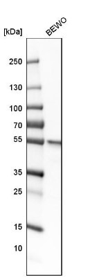
- Western Blot: LPCAT1 Antibody [NBP1-88923] - Analysis in human cell line BEWO.
- Submitted by
- Novus Biologicals (provider)
- Main image
- Experimental details
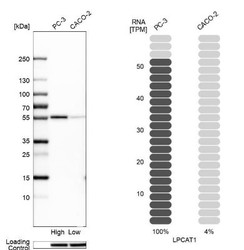
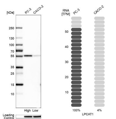
- Western Blot: LPCAT1 Antibody [NBP1-88923] - Analysis in human cell lines PC-3 and Caco-2 using anti-LPCAT1 antibody. Corresponding LPCAT1 RNA-seq data are presented for the same cell lines. Loading control: anti-HSP90B1.
Supportive validation
- Submitted by
- Novus Biologicals (provider)
- Main image
- Experimental details
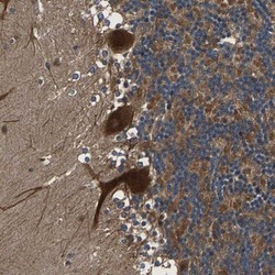
- Immunohistochemistry-Paraffin: LPCAT1 Antibody [NBP1-88923] - Staining of human cerebellum shows strong cytoplasmic positivity in Purkinje cells.
- Submitted by
- Novus Biologicals (provider)
- Main image
- Experimental details
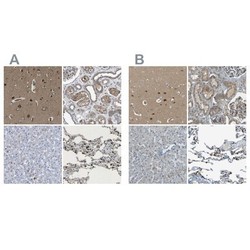
- Immunohistochemistry-Paraffin: LPCAT1 Antibody [NBP1-88923] - Staining of human cerebral cortex, kidney, liver and lung using Anti-LPCAT1 antibody NBP1-88923 (A) shows similar protein distribution across tissues to independent antibody NBP1-88922 (B).

- Submitted by
- Novus Biologicals (provider)
- Main image
- Experimental details
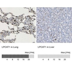
- Immunohistochemistry-Paraffin: LPCAT1 Antibody [NBP1-88923] - Analysis in human lung and liver tissues. Corresponding LPCAT1 RNA-seq data are presented for the same tissues.

- Submitted by
- Novus Biologicals (provider)
- Main image

- Experimental details
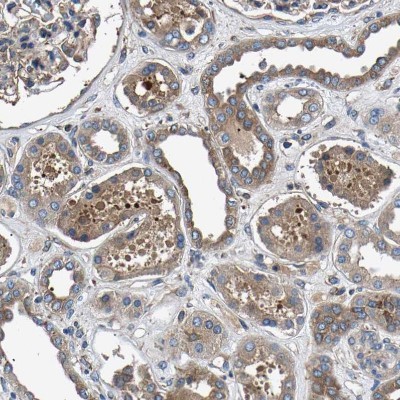
- Immunohistochemistry-Paraffin: LPCAT1 Antibody [NBP1-88923] - Staining of human kidney shows weak to moderate cytoplasmic positivity in cells in tubules.
- Submitted by
- Novus Biologicals (provider)
- Main image

- Experimental details
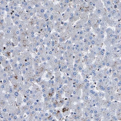
- Immunohistochemistry-Paraffin: LPCAT1 Antibody [NBP1-88923] - Staining of human liver shows no positivity in hepatocytes as expected.
- Submitted by
- Novus Biologicals (provider)
- Main image
- Experimental details
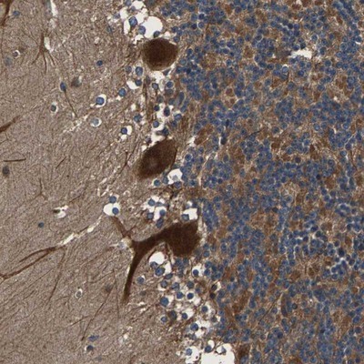
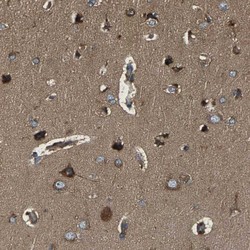
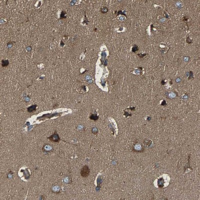
- Immunohistochemistry-Paraffin: LPCAT1 Antibody [NBP1-88923] - Staining of human cerebral cortex shows moderate to strong cytoplasmic positivity in neurons.
- Submitted by
- Novus Biologicals (provider)
- Main image
- Experimental details
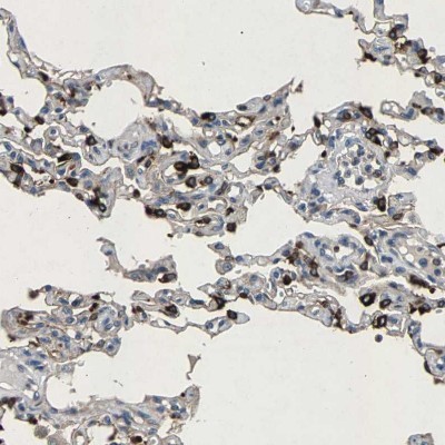
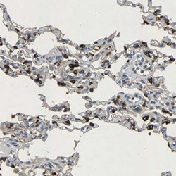
- Immunohistochemistry-Paraffin: LPCAT1 Antibody [NBP1-88923] - Staining of human lung shows moderate to strong cytoplasmic positivity in pneumocytes.