Explore
 Validate
 Learn
Learn Western blot
 Immunohistochemistry
Antibody data
- Antibody Data
- Antigen structure
- References [3]
- Comments [0]
- Validations
- Immunohistochemistry [1]
- Other assay [2]
Submit
Validation data
Reference
Comment
Report error
- Product number
- PA5-34801 - Provider product page

- Provider
- Invitrogen Antibodies
- Product name
- IL-21 Polyclonal Antibody
- Antibody type
- Polyclonal
- Antigen
- Recombinant full-length protein
- Description
- Recommended positive controls: HL-60. Predicted reactivity: Dog (80%), Pig (80%), Sheep (82%), Rhesus Monkey (96%), Bovine (81%). Store product as a concentrated solution. Centrifuge briefly prior to opening the vial.
- Reactivity
- Human
- Host
- Rabbit
- Isotype
- IgG
- Vial size
- 100 μL
- Concentration
- 1.02 mg/mL
- Storage
- Store at 4°C short term. For long term storage, store at -20°C, avoiding freeze/thaw cycles.
Submitted references Single-cell profiling of immune cells after neoadjuvant pembrolizumab and chemotherapy in IIIA non-small cell lung cancer (NSCLC).
Adenovirus-mediated overexpression of interleukin-21 regulates the development of oral squamous cell carcinoma in vitro.
Expression of the interleukin-21 and phosphorylated extracellular signal regulated kinase 1/2 in Kimura disease.
Hui Z, Zhang J, Ren Y, Li X, Yan C, Yu W, Wang T, Xiao S, Chen Y, Zhang R, Wei F, You J, Ren X
Cell death & disease 2022 Jul 13;13(7):607
Cell death & disease 2022 Jul 13;13(7):607
Adenovirus-mediated overexpression of interleukin-21 regulates the development of oral squamous cell carcinoma in vitro.
Liu H, Liu P, Sun D, Xing D, Wang X, Yang J, Wang S
Oncology letters 2020 Sep;20(3):3006-3014
Oncology letters 2020 Sep;20(3):3006-3014
Expression of the interleukin-21 and phosphorylated extracellular signal regulated kinase 1/2 in Kimura disease.
Chen QL, Li CX, Shao B, Gong ZC, Liu H, Ling B, Abasi K, Hu LL, Wang B, Yin XP
Journal of clinical pathology 2017 Aug;70(8):684-689
Journal of clinical pathology 2017 Aug;70(8):684-689
No comments: Submit comment
Supportive validation
- Submitted by
- Invitrogen Antibodies (provider)
- Main image
- Experimental details
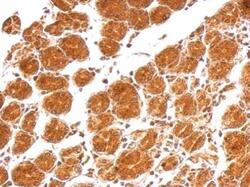
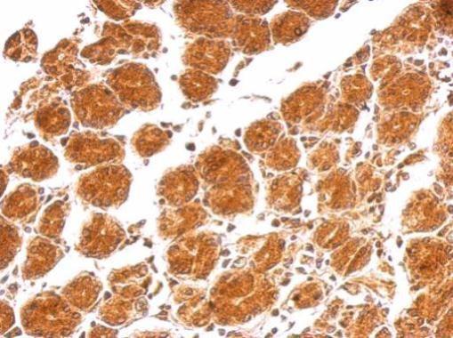
- IL-21 Polyclonal Antibody detects IL21 protein at cytosol on human colon carcinoma by immunohistochemical analysis. Sample: Paraffin-embedded colon carcinoma. IL-21 Polyclonal Antibody (Product # PA5-34801) dilution: 1:500. Antigen Retrieval: EDTA based buffer, pH 8.0, 15 min.
Supportive validation
- Submitted by
- Invitrogen Antibodies (provider)
- Main image
- Experimental details
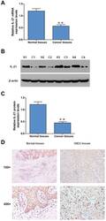
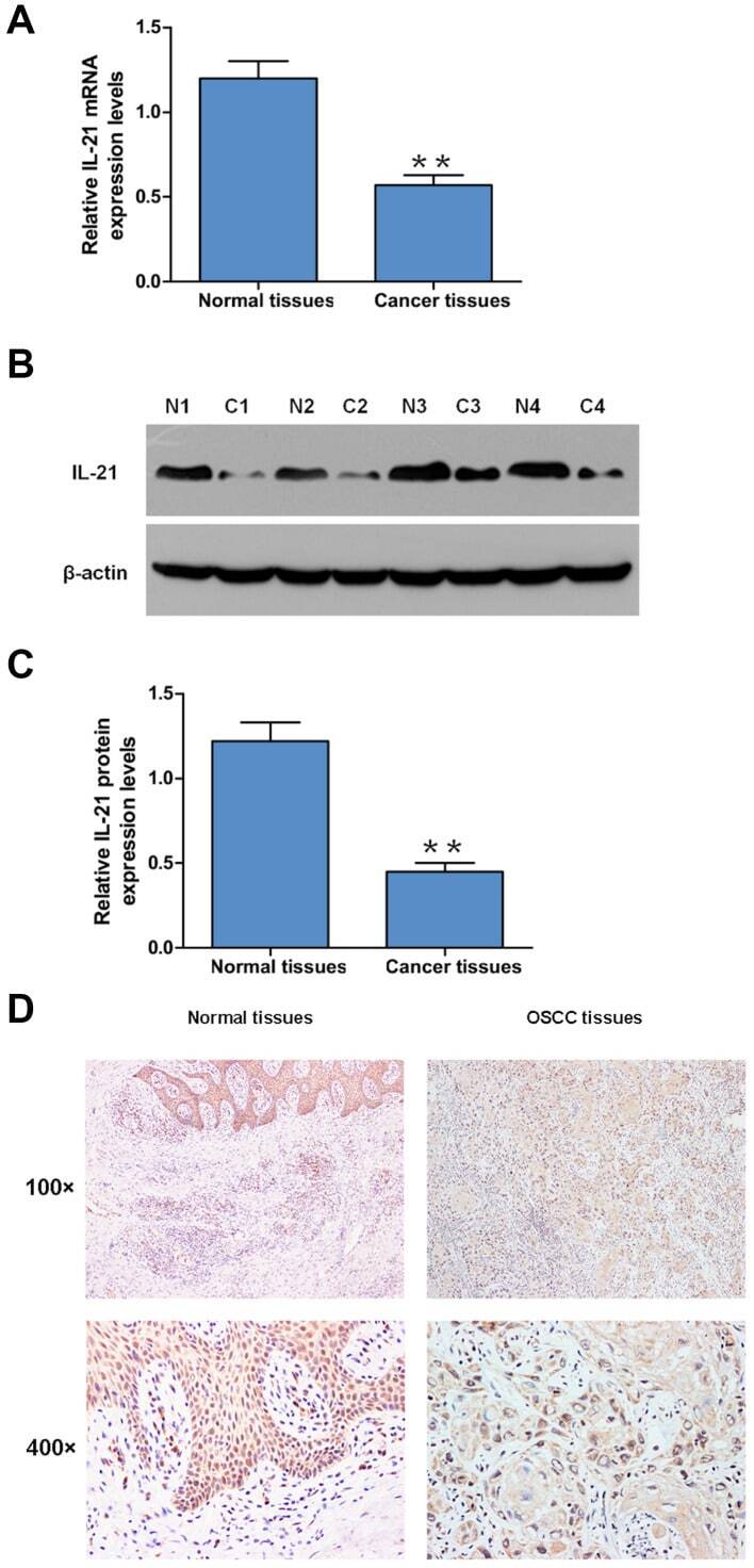
- Figure 1. Expression levels of IL-21 in OSCC tissues. (A) mRNA expression levels of IL-21 in OSCC tissues and adjacent normal tissues were detected using reverse transcription-quantitative PCR. (B) Protein expression levels of IL-21 in OSCC tissues and adjacent normal tissues were detected using western blotting. (C) Semi-quantification of the western blotting data presented in part B. Expression levels of IL-21 were normalized to beta-actin. The data are presented as the mean +- SEM from three independent experiments. **P
- Submitted by
- Invitrogen Antibodies (provider)
- Main image
- Experimental details
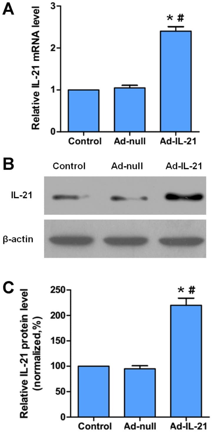
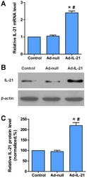
- Figure 2. Adenoviral-mediated overexpression of IL-21 in CAL-27 cells. (A) Reverse transcription-quantitative PCR or (B) western blotting was used to analyze the mRNA or protein expression levels, respectively, of IL-21 in CAL-27 cells infected with Ad- or Ad-IL-21. (C) Semi-quantification of the western blotting data presented in part B. Expression levels of IL-21 were normalized to beta-actin. The data were expressed as the mean +- SEM from three independent experiments. *P