Explore
 Validate
 Learn
Learn ELISA
ELISAAntibody data
- Antibody Data
- Antigen structure
- References [9]
- Comments [0]
- Validations
- ELISA [1]
- Other assay [6]
Submit
Validation data
Reference
Comment
Report error
- Product number
- 14-7219-81 - Provider product page

- Provider
- Invitrogen Antibodies
- Product name
- IL-21 Monoclonal Antibody (eBio3A3-N2 (3A3-N2)), eBioscience™
- Antibody type
- Monoclonal
- Antigen
- Other
- Description
- Description: The monoclonal antibody eBio3A3-N2 reacts with human IL-21, a potent stimulator of NK and cytolytic T cells. IL-21 is a 131 amino acid protein most closely related to IL-2 and IL-15. Expression is found in CD4+ cells upon activation. In addition to its effect on NK cells, IL-21 stimulates proliferation of B-cell stimulated by crosslinking of the CD40 antigen and stimulates the proliferation of bone marrow progenitor cells and the expression of the NK-cell marker CD56 in the presence of IL-15. Applications Reported: The eBio3A3-N2 antibody has been reported for use in ELISA capture of IL-21 analysis. Applications Tested: The eBio3A3-N2 antibody has been tested as the capture antibody in a sandwich ELISA for analysis of human Interleukin-21 (IL-21) in combination with the biotin eBio2B2-G20 (Product # 13-7218-81) antibody for detection and recombinant standard human IL-21 (Product # 39-8219-65) as the standard. A suitable range of concentrations of this antibody for ELISA capture is 1-4 µg/mL. A standard curve consisting of doubling dilutions of the recombinant standard over the range of 4000 pg/mL - 60 pg/mL should be included in each ELISA plate. Purity: Greater than 90%, as determined by SDS-PAGE. Aggregation: Less than 10%, as determined by HPLC. Filtration: 0.2 µm post-manufacturing filtered.
- Reactivity
- Human
- Host
- Mouse
- Isotype
- IgG
- Antibody clone number
- eBio3A3-N2 (3A3-N2)
- Vial size
- 50 µg
- Concentration
- 0.5 mg/mL
- Storage
- 4° C
Submitted references Ageing promotes early T follicular helper cell differentiation by modulating expression of RBPJ.
Effects of Anti-TNFα Treatment on Mucosal Expression of IL-17A, IL-21, and IL-22 and Cytokine-Producing T Cell Subsets in Crohn's Disease.
C5aR1 regulates T follicular helper differentiation and chronic graft-versus-host disease bronchiolitis obliterans.
Overexpression of microRNA-155 increases IL-21 mediated STAT3 signaling and IL-21 production in systemic lupus erythematosus.
Circulating CXCR5+PD-1+ response predicts influenza vaccine antibody responses in young adults but not elderly adults.
Reprogramming CD19-specific T cells with IL-21 signaling can improve adoptive immunotherapy of B-lineage malignancies.
Characterization of interleukin-17-producing regulatory T cells in inflamed intestinal mucosa from patients with inflammatory bowel diseases.
Interleukin-21 and the IL-21 receptor: novel effectors of NK and T cell responses.
Interleukin 21 and its receptor are involved in NK cell expansion and regulation of lymphocyte function.
Webb LMC, Fra-Bido S, Innocentin S, Matheson LS, Attaf N, Bignon A, Novarino J, Fazilleau N, Linterman MA
Aging cell 2021 Jan;20(1):e13295
Aging cell 2021 Jan;20(1):e13295
Effects of Anti-TNFα Treatment on Mucosal Expression of IL-17A, IL-21, and IL-22 and Cytokine-Producing T Cell Subsets in Crohn's Disease.
Dige A, Magnusson MK, Uhrenholt C, Rasmussen TK, Kragstrup T, Öhman L, Dahlerup J, Agnholt J
Mediators of inflammation 2018;2018:3279607
Mediators of inflammation 2018;2018:3279607
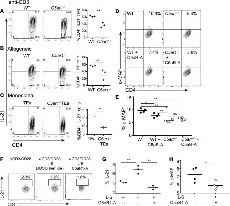
C5aR1 regulates T follicular helper differentiation and chronic graft-versus-host disease bronchiolitis obliterans.
Verghese DA, Chun N, Paz K, Fribourg M, Woodruff TM, Flynn R, Hu Y, Xiong H, Zhang W, Yi Z, Du J, Blazar BR, Heeger PS
JCI insight 2018 Dec 20;3(24)
JCI insight 2018 Dec 20;3(24)
Overexpression of microRNA-155 increases IL-21 mediated STAT3 signaling and IL-21 production in systemic lupus erythematosus.
Rasmussen TK, Andersen T, Bak RO, Yiu G, Sørensen CM, Stengaard-Pedersen K, Mikkelsen JG, Utz PJ, Holm CK, Deleuran B
Arthritis research & therapy 2015 Jun 9;17(1):154
Arthritis research & therapy 2015 Jun 9;17(1):154
Circulating CXCR5+PD-1+ response predicts influenza vaccine antibody responses in young adults but not elderly adults.
Herati RS, Reuter MA, Dolfi DV, Mansfield KD, Aung H, Badwan OZ, Kurupati RK, Kannan S, Ertl H, Schmader KE, Betts MR, Canaday DH, Wherry EJ
Journal of immunology (Baltimore, Md. : 1950) 2014 Oct 1;193(7):3528-37
Journal of immunology (Baltimore, Md. : 1950) 2014 Oct 1;193(7):3528-37
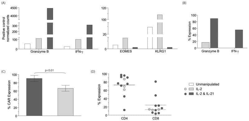
Reprogramming CD19-specific T cells with IL-21 signaling can improve adoptive immunotherapy of B-lineage malignancies.
Singh H, Figliola MJ, Dawson MJ, Huls H, Olivares S, Switzer K, Mi T, Maiti S, Kebriaei P, Lee DA, Champlin RE, Cooper LJ
Cancer research 2011 May 15;71(10):3516-27
Cancer research 2011 May 15;71(10):3516-27
Characterization of interleukin-17-producing regulatory T cells in inflamed intestinal mucosa from patients with inflammatory bowel diseases.
Hovhannisyan Z, Treatman J, Littman DR, Mayer L
Gastroenterology 2011 Mar;140(3):957-65
Gastroenterology 2011 Mar;140(3):957-65
Interleukin-21 and the IL-21 receptor: novel effectors of NK and T cell responses.
Parrish-Novak J, Foster DC, Holly RD, Clegg CH
Journal of leukocyte biology 2002 Nov;72(5):856-63
Journal of leukocyte biology 2002 Nov;72(5):856-63
Interleukin 21 and its receptor are involved in NK cell expansion and regulation of lymphocyte function.
Parrish-Novak J, Dillon SR, Nelson A, Hammond A, Sprecher C, Gross JA, Johnston J, Madden K, Xu W, West J, Schrader S, Burkhead S, Heipel M, Brandt C, Kuijper JL, Kramer J, Conklin D, Presnell SR, Berry J, Shiota F, Bort S, Hambly K, Mudri S, Clegg C, Moore M, Grant FJ, Lofton-Day C, Gilbert T, Rayond F, Ching A, Yao L, Smith D, Webster P, Whitmore T, Maurer M, Kaushansky K, Holly RD, Foster D
Nature 2000 Nov 2;408(6808):57-63
Nature 2000 Nov 2;408(6808):57-63
No comments: Submit comment
Supportive validation
- Submitted by
- Invitrogen Antibodies (provider)
- Main image

- Experimental details
- Standard curve of human IL-21 ELISA.

Supportive validation
- Submitted by
- Invitrogen Antibodies (provider)
- Main image

- Experimental details
- NULL
- Submitted by
- Invitrogen Antibodies (provider)
- Main image

- Experimental details
- NULL
- Submitted by
- Invitrogen Antibodies (provider)
- Main image
- Experimental details
- NULL

- Submitted by
- Invitrogen Antibodies (provider)
- Main image
- Experimental details
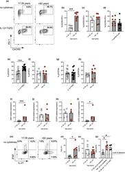
- FIGURE 2 Age promotes pre-Tfh cell differentiation in humans. Flow plots showing the frequency of CXCR5 + PD-1 + cells amongst CD4 + following 4 days in vitro activation of naive CD4 + T cells taken from younger (17-39 yrs) and older (>60 yrs) donors in the presence/absence IL-12 and TGFbeta (a). Percentage of CXCR5 + PD-1 + following activation of naive CD4 + T cells from younger and older donors in the absence (b) or presence (c) of IL-12 and TGFbeta. Percentage of CXCR5 + PD1 + cells following activation of naive CD4 + T cells from older donors with or without neutralising antibodies to IL-12, Activin A and TGFbeta (d). Bar graphs showing percentage of cells expressing pSTAT3 (e, f), and pSTAT5 (g, h) on day 3 of culture in presence/absence of IL-12 and TGFbeta in donors of the indicated ages. RT-PCR determination MAF (i), BCL6 (j), FOXP3 (k) and CXCL13 (l) following 4 days activation of naive CD4 + T cells from younger and older donors in the absence of polarising cytokines. Flow plots (m-o) and bar graphs (n-o) showing percentage of IFNgamma and IL-21 expressing cells following PMA and ionomycin mediated restimulation after 4 days in vitro culture of naive CD4 + T cells from younger and older donors in the absence of IL-12 and TGFbeta (m-o). Bar graph showing levels of IgG produced by B cells following co-culture with CD4+ T cells from day four cultures from young and older donors, in the presence/absence of Tfh-polarising cytokines (p). Each symbol is representative of
- Submitted by
- Invitrogen Antibodies (provider)
- Main image
- Experimental details
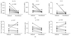
- Figure 1 Mucosal gene expression and LP T cell producing IL-17A, IL-21, and IL-22 at baseline in inflamed and noninflamed mucosa. Gene expression was determined by rtPCR (a) and frequencies of IL-17A-, IL-21-, and IL-22-producing cells among LP CD3 + T cells were determined by flow cytometry (b). Gene expression data is displayed as the normalized ratios between the relative expression of the gene of interest and the housekeeping gene HPRT-1 . Wilcoxon signed-rank test for comparison was applied on paired samples (rtPCR: IL-17A n = 11; IL-21 n = 9; IL-22 n = 9; flow cytometry: n = 8 for each cytokine).
- Submitted by
- Invitrogen Antibodies (provider)
- Main image
- Experimental details
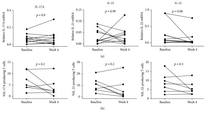
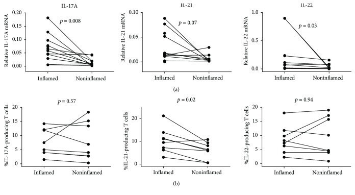
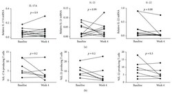
- Figure 2 Comparison of mucosal gene expression and LP T cells expressing IL-17A, IL-21, and IL-22 between baseline and week 4 of adalimumab treatment. Gene expression was determined by rtPCR (a) and frequencies of IL-17A-, IL-21-, and IL-22-producing cells among LP CD3 + T cells were determined by flow cytometry (b). Gene expression data is displayed as the normalized ratios between the relative expression of the gene of interest and the housekeeping gene HPRT-1 . Wilcoxon signed-rank test for comparison was applied on paired samples (rtPCR: IL-17A n = 12; IL-21 n = 11; IL-22 n = 11; flow cytometry: n = 7 for each cytokine).