Explore
 Validate
 Learn
Learn Flow cytometry
Antibody data
- Antibody Data
- Antigen structure
- References [20]
- Comments [0]
- Validations
- Flow cytometry [2]
- Other assay [6]
Submit
Validation data
Reference
Comment
Report error
- Product number
- 12-7219-41 - Provider product page

- Provider
- Invitrogen Antibodies
- Product name
- IL-21 Monoclonal Antibody (eBio3A3-N2 (3A3-N2)), PE, eBioscience™
- Antibody type
- Monoclonal
- Antigen
- Other
- Description
- Description: The monoclonal antibody eBio3A3-N2 reacts with human IL-21, a potent stimulator of NK and cytolytic T cells. IL-21 is a 131 amino acid protein most closely related to IL-2 and IL-15. Expression is found in CD4+ cells upon activation. In addition to its effect on NK cells, IL-21 stimulates proliferation of B-cell stimulated by crosslinking of the CD40 antigen and stimulates the proliferation of bone marrow progenitor cells and the expression of the NK-cell marker CD56 in the presence of IL-15. Applications Reported: This eBio3A3-N2 (3A3-N2) antibody has been reported for use in intracellular staining followed by flow cytometric analysis. Applications Tested: This eBio3A3-N2 (3A3-N2) antibody has been pre-titrated and tested by intracellular staining of restimulated, Th17-polarized normal human peripheral blood cells. This can be used at 5 µL (0.5 µg) per test. A test is defined as the amount (µg) of antibody that will stain a cell sample in a final volume of 100 µL. Cell number should be determined empirically but can range from 10^5 to 10^8 cells/test. Staining for IL-21 has shown donor variability. stimulation times may vary depending on the donor and should be optimized. Increased staining levels have been observed on Th17-polarized normal human peripheral blood cells. Excitation: 488-561 nm; Emission: 578 nm; Laser: Blue Laser, Green Laser, Yellow-Green Laser. Filtration: 0.2 µm post-manufacturing filtered.
- Reactivity
- Human
- Host
- Mouse
- Conjugate
- Yellow dye
- Isotype
- IgG
- Antibody clone number
- eBio3A3-N2 (3A3-N2)
- Vial size
- 25 Tests
- Concentration
- 5 μL/Test
- Storage
- 4°C, store in dark, DO NOT FREEZE!
Submitted references Human IRF1 governs macrophagic IFN-γ immunity to mycobacteria.
Ageing promotes early T follicular helper cell differentiation by modulating expression of RBPJ.
Functional SARS-CoV-2-Specific Immune Memory Persists after Mild COVID-19.
A delayed fractionated dose RTS,S AS01 vaccine regimen mediates protection via improved T follicular helper and B cell responses.
Effects of Anti-TNFα Treatment on Mucosal Expression of IL-17A, IL-21, and IL-22 and Cytokine-Producing T Cell Subsets in Crohn's Disease.
Genetic Architecture of Adaptive Immune System Identifies Key Immune Regulators.
Hepatocyte-derived exosomes promote T follicular regulatory cell expansion during hepatitis C virus infection.
Activated T follicular helper-like cells are released into blood after oral vaccination and correlate with vaccine specific mucosal B-cell memory.
C5aR1 regulates T follicular helper differentiation and chronic graft-versus-host disease bronchiolitis obliterans.
TIGIT expressing CD4+T cells represent a tumor-supportive T cell subset in chronic lymphocytic leukemia.
Th1 is the predominant helper T cell subset that produces GM-CSF in the joint of rheumatoid arthritis.
Cytotoxic T Cell Functions Accumulate When CD4 Is Downregulated by CD4(+) T Cells in African Green Monkeys.
Overexpression of microRNA-155 increases IL-21 mediated STAT3 signaling and IL-21 production in systemic lupus erythematosus.
Impaired Phenotype and Function of T Follicular Helper Cells in HIV-1-Infected Children Receiving ART.
Galectin-9 and IL-21 mediate cross-regulation between Th17 and Treg cells during acute hepatitis C.
Increased frequency of circulating IL-21 producing Th-cells in patients with granulomatosis with polyangiitis (GPA).
Engineering lymph node homing of ex vivo-expanded human natural killer cells via trogocytosis of the chemokine receptor CCR7.
Increase in IL-21 producing T-cells in patients with systemic lupus erythematosus.
Reprogramming CD19-specific T cells with IL-21 signaling can improve adoptive immunotherapy of B-lineage malignancies.
1,25-Dihydroxyvitamin D3 and IL-2 combine to inhibit T cell production of inflammatory cytokines and promote development of regulatory T cells expressing CTLA-4 and FoxP3.
Rosain J, Neehus AL, Manry J, Yang R, Le Pen J, Daher W, Liu Z, Chan YH, Tahuil N, Türel Ö, Bourgey M, Ogishi M, Doisne JM, Izquierdo HM, Shirasaki T, Le Voyer T, Guérin A, Bastard P, Moncada-Vélez M, Han JE, Khan T, Rapaport F, Hong SH, Cheung A, Haake K, Mindt BC, Pérez L, Philippot Q, Lee D, Zhang P, Rinchai D, Al Ali F, Ahmad Ata MM, Rahman M, Peel JN, Heissel S, Molina H, Kendir-Demirkol Y, Bailey R, Zhao S, Bohlen J, Mancini M, Seeleuthner Y, Roelens M, Lorenzo L, Soudée C, Paz MEJ, González ML, Jeljeli M, Soulier J, Romana S, L'Honneur AS, Materna M, Martínez-Barricarte R, Pochon M, Oleaga-Quintas C, Michev A, Migaud M, Lévy R, Alyanakian MA, Rozenberg F, Croft CA, Vogt G, Emile JF, Kremer L, Ma CS, Fritz JH, Lemon SM, Spaan AN, Manel N, Abel L, MacDonald MR, Boisson-Dupuis S, Marr N, Tangye SG, Di Santo JP, Zhang Q, Zhang SY, Rice CM, Béziat V, Lachmann N, Langlais D, Casanova JL, Gros P, Bustamante J
Cell 2023 Feb 2;186(3):621-645.e33
Cell 2023 Feb 2;186(3):621-645.e33
Ageing promotes early T follicular helper cell differentiation by modulating expression of RBPJ.
Webb LMC, Fra-Bido S, Innocentin S, Matheson LS, Attaf N, Bignon A, Novarino J, Fazilleau N, Linterman MA
Aging cell 2021 Jan;20(1):e13295
Aging cell 2021 Jan;20(1):e13295
Functional SARS-CoV-2-Specific Immune Memory Persists after Mild COVID-19.
Rodda LB, Netland J, Shehata L, Pruner KB, Morawski PA, Thouvenel CD, Takehara KK, Eggenberger J, Hemann EA, Waterman HR, Fahning ML, Chen Y, Hale M, Rathe J, Stokes C, Wrenn S, Fiala B, Carter L, Hamerman JA, King NP, Gale M Jr, Campbell DJ, Rawlings DJ, Pepper M
Cell 2021 Jan 7;184(1):169-183.e17
Cell 2021 Jan 7;184(1):169-183.e17
A delayed fractionated dose RTS,S AS01 vaccine regimen mediates protection via improved T follicular helper and B cell responses.
Pallikkuth S, Chaudhury S, Lu P, Pan L, Jongert E, Wille-Reece U, Pahwa S
eLife 2020 Apr 29;9
eLife 2020 Apr 29;9
Effects of Anti-TNFα Treatment on Mucosal Expression of IL-17A, IL-21, and IL-22 and Cytokine-Producing T Cell Subsets in Crohn's Disease.
Dige A, Magnusson MK, Uhrenholt C, Rasmussen TK, Kragstrup T, Öhman L, Dahlerup J, Agnholt J
Mediators of inflammation 2018;2018:3279607
Mediators of inflammation 2018;2018:3279607
Genetic Architecture of Adaptive Immune System Identifies Key Immune Regulators.
Lagou V, Garcia-Perez JE, Smets I, Van Horebeek L, Vandebergh M, Chen L, Mallants K, Prezzemolo T, Hilven K, Humblet-Baron S, Moisse M, Van Damme P, Boeckxstaens G, Bowness P, Dubois B, Dooley J, Liston A, Goris A
Cell reports 2018 Oct 16;25(3):798-810.e6
Cell reports 2018 Oct 16;25(3):798-810.e6
Hepatocyte-derived exosomes promote T follicular regulatory cell expansion during hepatitis C virus infection.
Cobb DA, Kim OK, Golden-Mason L, Rosen HR, Hahn YS
Hepatology (Baltimore, Md.) 2018 Jan;67(1):71-85
Hepatology (Baltimore, Md.) 2018 Jan;67(1):71-85
Activated T follicular helper-like cells are released into blood after oral vaccination and correlate with vaccine specific mucosal B-cell memory.
Cárdeno A, Magnusson MK, Quiding-Järbrink M, Lundgren A
Scientific reports 2018 Feb 9;8(1):2729
Scientific reports 2018 Feb 9;8(1):2729
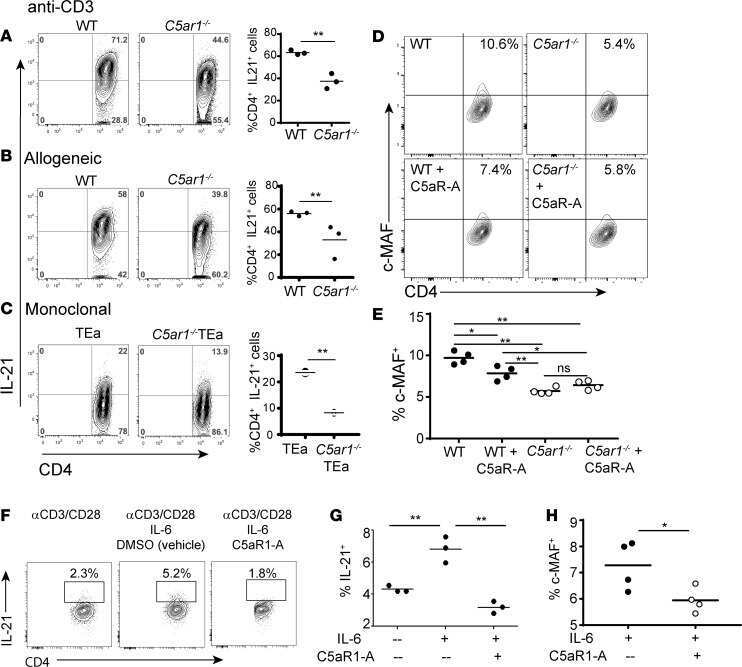
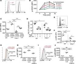
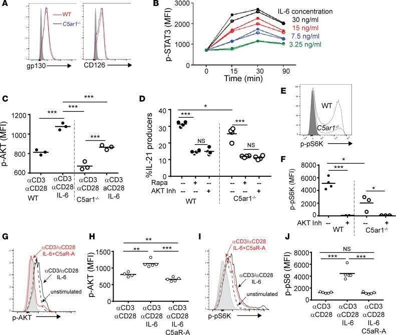
C5aR1 regulates T follicular helper differentiation and chronic graft-versus-host disease bronchiolitis obliterans.
Verghese DA, Chun N, Paz K, Fribourg M, Woodruff TM, Flynn R, Hu Y, Xiong H, Zhang W, Yi Z, Du J, Blazar BR, Heeger PS
JCI insight 2018 Dec 20;3(24)
JCI insight 2018 Dec 20;3(24)
TIGIT expressing CD4+T cells represent a tumor-supportive T cell subset in chronic lymphocytic leukemia.
Catakovic K, Gassner FJ, Ratswohl C, Zaborsky N, Rebhandl S, Schubert M, Steiner M, Gutjahr JC, Pleyer L, Egle A, Hartmann TN, Greil R, Geisberger R
Oncoimmunology 2017;7(1):e1371399
Oncoimmunology 2017;7(1):e1371399
Th1 is the predominant helper T cell subset that produces GM-CSF in the joint of rheumatoid arthritis.
Yamada H, Haraguchi A, Sakuraba K, Okazaki K, Fukushi JI, Mizu-Uchi H, Akasaki Y, Esaki Y, Kamura S, Fujimura K, Kondo M, Miyahara H, Nakashima Y, Yoshikai Y
RMD open 2017;3(1):e000487
RMD open 2017;3(1):e000487
Cytotoxic T Cell Functions Accumulate When CD4 Is Downregulated by CD4(+) T Cells in African Green Monkeys.
Vinton CL, Ortiz AM, Calantone N, Mudd JC, Deleage C, Morcock DR, Whitted S, Estes JD, Hirsch VM, Brenchley JM
Journal of immunology (Baltimore, Md. : 1950) 2017 Jun 1;198(11):4403-4412
Journal of immunology (Baltimore, Md. : 1950) 2017 Jun 1;198(11):4403-4412
Overexpression of microRNA-155 increases IL-21 mediated STAT3 signaling and IL-21 production in systemic lupus erythematosus.
Rasmussen TK, Andersen T, Bak RO, Yiu G, Sørensen CM, Stengaard-Pedersen K, Mikkelsen JG, Utz PJ, Holm CK, Deleuran B
Arthritis research & therapy 2015 Jun 9;17(1):154
Arthritis research & therapy 2015 Jun 9;17(1):154
Impaired Phenotype and Function of T Follicular Helper Cells in HIV-1-Infected Children Receiving ART.
Bekele Y, Amu S, Bobosha K, Lantto R, Nilsson A, Endale B, Gebre M, Aseffa A, Rethi B, Howe R, Chiodi F
Medicine 2015 Jul;94(27):e1125
Medicine 2015 Jul;94(27):e1125
Galectin-9 and IL-21 mediate cross-regulation between Th17 and Treg cells during acute hepatitis C.
Kared H, Fabre T, Bédard N, Bruneau J, Shoukry NH
PLoS pathogens 2013;9(6):e1003422
PLoS pathogens 2013;9(6):e1003422
Increased frequency of circulating IL-21 producing Th-cells in patients with granulomatosis with polyangiitis (GPA).
Abdulahad WH, Lepse N, Stegeman CA, Huitema MG, Doornbos-van der Meer B, Tadema H, Rutgers A, Limburg PC, Kallenberg CG, Heeringa P
Arthritis research & therapy 2013;15(3):R70
Arthritis research & therapy 2013;15(3):R70
Engineering lymph node homing of ex vivo-expanded human natural killer cells via trogocytosis of the chemokine receptor CCR7.
Somanchi SS, Somanchi A, Cooper LJ, Lee DA
Blood 2012 May 31;119(22):5164-72
Blood 2012 May 31;119(22):5164-72
Increase in IL-21 producing T-cells in patients with systemic lupus erythematosus.
Dolff S, Abdulahad WH, Westra J, Doornbos-van der Meer B, Limburg PC, Kallenberg CG, Bijl M
Arthritis research & therapy 2011;13(5):R157
Arthritis research & therapy 2011;13(5):R157
Reprogramming CD19-specific T cells with IL-21 signaling can improve adoptive immunotherapy of B-lineage malignancies.
Singh H, Figliola MJ, Dawson MJ, Huls H, Olivares S, Switzer K, Mi T, Maiti S, Kebriaei P, Lee DA, Champlin RE, Cooper LJ
Cancer research 2011 May 15;71(10):3516-27
Cancer research 2011 May 15;71(10):3516-27
1,25-Dihydroxyvitamin D3 and IL-2 combine to inhibit T cell production of inflammatory cytokines and promote development of regulatory T cells expressing CTLA-4 and FoxP3.
Jeffery LE, Burke F, Mura M, Zheng Y, Qureshi OS, Hewison M, Walker LS, Lammas DA, Raza K, Sansom DM
Journal of immunology (Baltimore, Md. : 1950) 2009 Nov 1;183(9):5458-67
Journal of immunology (Baltimore, Md. : 1950) 2009 Nov 1;183(9):5458-67
No comments: Submit comment
Supportive validation
- Submitted by
- Invitrogen Antibodies (provider)
- Main image
- Experimental details
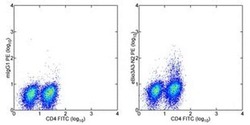
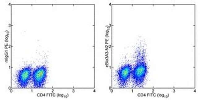
- Intracellular staining of normal human peripheral blood cells treated with PMA and Ionomycin in the presence of Brefeldin A for 5 hours with Anti-Human CD4 FITC (Product # 11-0049-42) and Mouse IgG1 kappa Isotype Control PE (Product # 12-4714-42) (left) or Anti-Human IL-21 PE (right). Cells in the lymphocyte gate were used for analysis.
- Conjugate
- Yellow dye
- Submitted by
- Invitrogen Antibodies (provider)
- Main image

- Experimental details
- Intracellular staining of normal human peripheral blood cells treated with PMA and Ionomycin in the presence of Brefeldin A for 5 hours with Anti-Human CD4 FITC (Product # 11-0049-42) and Mouse IgG1 kappa Isotype Control PE (Product # 12-4714-42) (left) or Anti-Human IL-21 PE (right). Cells in the lymphocyte gate were used for analysis.

Supportive validation
- Submitted by
- Invitrogen Antibodies (provider)
- Main image
- Experimental details
- NULL
- Conjugate
- Yellow dye

- Submitted by
- Invitrogen Antibodies (provider)
- Main image

- Experimental details
- NULL
- Conjugate
- Yellow dye
- Submitted by
- Invitrogen Antibodies (provider)
- Main image
- Experimental details
- NULL
- Conjugate
- Yellow dye
- Submitted by
- Invitrogen Antibodies (provider)
- Main image
- Experimental details
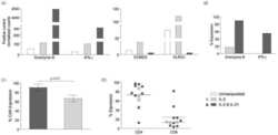
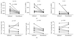
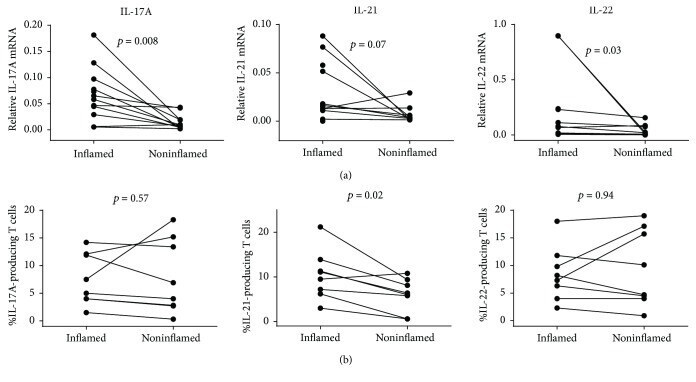
- Figure 1 Mucosal gene expression and LP T cell producing IL-17A, IL-21, and IL-22 at baseline in inflamed and noninflamed mucosa. Gene expression was determined by rtPCR (a) and frequencies of IL-17A-, IL-21-, and IL-22-producing cells among LP CD3 + T cells were determined by flow cytometry (b). Gene expression data is displayed as the normalized ratios between the relative expression of the gene of interest and the housekeeping gene HPRT-1 . Wilcoxon signed-rank test for comparison was applied on paired samples (rtPCR: IL-17A n = 11; IL-21 n = 9; IL-22 n = 9; flow cytometry: n = 8 for each cytokine).
- Conjugate
- Yellow dye
- Submitted by
- Invitrogen Antibodies (provider)
- Main image
- Experimental details
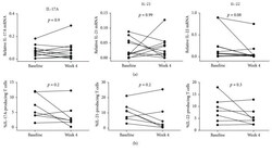
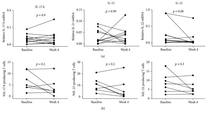
- Figure 2 Comparison of mucosal gene expression and LP T cells expressing IL-17A, IL-21, and IL-22 between baseline and week 4 of adalimumab treatment. Gene expression was determined by rtPCR (a) and frequencies of IL-17A-, IL-21-, and IL-22-producing cells among LP CD3 + T cells were determined by flow cytometry (b). Gene expression data is displayed as the normalized ratios between the relative expression of the gene of interest and the housekeeping gene HPRT-1 . Wilcoxon signed-rank test for comparison was applied on paired samples (rtPCR: IL-17A n = 12; IL-21 n = 11; IL-22 n = 11; flow cytometry: n = 7 for each cytokine).
- Conjugate
- Yellow dye
- Submitted by
- Invitrogen Antibodies (provider)
- Main image
- Experimental details
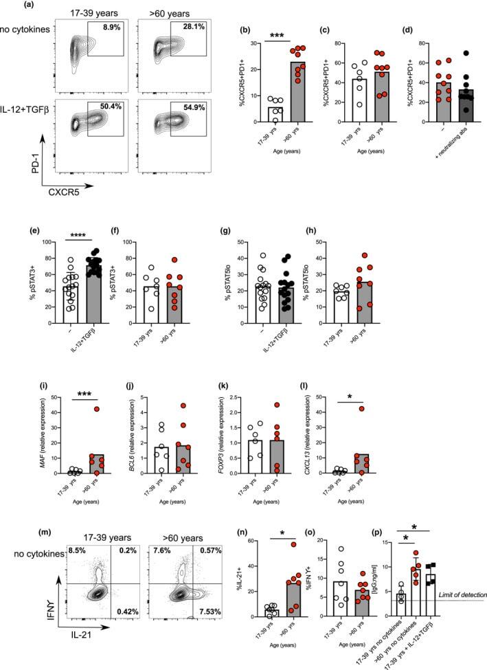
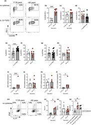
- FIGURE 2 Age promotes pre-Tfh cell differentiation in humans. Flow plots showing the frequency of CXCR5 + PD-1 + cells amongst CD4 + following 4 days in vitro activation of naive CD4 + T cells taken from younger (17-39 yrs) and older (>60 yrs) donors in the presence/absence IL-12 and TGFbeta (a). Percentage of CXCR5 + PD-1 + following activation of naive CD4 + T cells from younger and older donors in the absence (b) or presence (c) of IL-12 and TGFbeta. Percentage of CXCR5 + PD1 + cells following activation of naive CD4 + T cells from older donors with or without neutralising antibodies to IL-12, Activin A and TGFbeta (d). Bar graphs showing percentage of cells expressing pSTAT3 (e, f), and pSTAT5 (g, h) on day 3 of culture in presence/absence of IL-12 and TGFbeta in donors of the indicated ages. RT-PCR determination MAF (i), BCL6 (j), FOXP3 (k) and CXCL13 (l) following 4 days activation of naive CD4 + T cells from younger and older donors in the absence of polarising cytokines. Flow plots (m-o) and bar graphs (n-o) showing percentage of IFNgamma and IL-21 expressing cells following PMA and ionomycin mediated restimulation after 4 days in vitro culture of naive CD4 + T cells from younger and older donors in the absence of IL-12 and TGFbeta (m-o). Bar graph showing levels of IgG produced by B cells following co-culture with CD4+ T cells from day four cultures from young and older donors, in the presence/absence of Tfh-polarising cytokines (p). Each symbol is representative of
- Conjugate
- Yellow dye