 Explore
 Validate
 Learn
Learn Western blot
Antibody data
- Antibody Data
- Antigen structure
- References [1]
- Comments [0]
- Validations
- Western blot [2]
- Immunocytochemistry [1]
- Other assay [1]
Submit
Validation data
Reference
Comment
Report error
- Product number
- PA1-24931 - Provider product page

- Provider
- Invitrogen Antibodies
- Product name
- Cofilin Polyclonal Antibody
- Antibody type
- Polyclonal
- Antigen
- Synthetic peptide
- Description
- Recommended positive controls: A431, PC-12, NIH3T3, MDCK.
- Concentration
- 11.4 mg/mL
Submitted references Cyclosporine A inhibits MRTF-SRF signaling through Na(+)/K(+) ATPase inhibition and actin remodeling.
Burat B, Faucher Q, Čechová P, Arnion H, Di Meo F, Sauvage FL, Marquet P, Essig M
FASEB bioAdvances 2019 Sep;1(9):561-578
FASEB bioAdvances 2019 Sep;1(9):561-578
No comments: Submit comment
Supportive validation
- Submitted by
- Invitrogen Antibodies (provider)
- Main image
- Experimental details
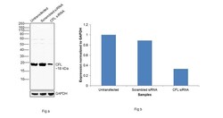
- Knockdown of Cofilin was achieved by transfecting HeLa with Cofilin specific siRNAs (Silencer® select Product # s2037, s2936 ). Western blot analysis (Fig. a) was performed using Whole cell extracts from the untransfected cells (lane 1), non-targeting scrambled siRNA transfected cells (lane 2) and,Cofilin knockdown cells (lane 3). The blot was probed with Cofilin Polyclonal Antibody (Product # PA1-24931, 1:10000 dilution) and Goat anti-Rabbit IgG (H+L) Superclonal™ Recombinant Secondary Antibody, HRP (Product # A27036, 1:4000 dilution). Densitometric analysis of this western blot is shown in histogram (Fig. b). Decrease in signal upon siRNA mediated knock down confirms that antibody is specific to Cofilin.

- Submitted by
- Invitrogen Antibodies (provider)
- Main image
- Experimental details
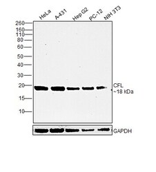
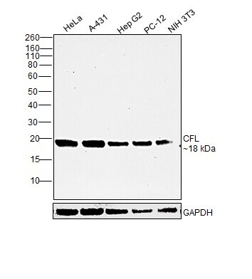
- Western blot was performed using Anti-Cofilin Polyclonal Antibody(Product # PA1-24931) and an 18kDa band corresponding to Cofilin was observed across cell lines tested. Whole cell extracts (30 µg lysate) of HeLa (Lane 1), A-431 (Lane 2), Hep G2 (Lane 3), PC-12 (Lane 4) and NIH/3T3 (Lane 5) were electrophoresed using NuPAGE™ 12% Bis-Tris Protein Gel (Product # NP0341BOX). Resolved proteins were then transferred onto a Nitrocellulose membrane (Product # IB23001) by iBlot® 2 Dry Blotting System (Product # IB21001). The blot was probed with the primary antibody (1:10000 dilution) and detected by chemiluminescence with Goat anti-Rabbit IgG (H+L) Superclonal™ Recombinant Secondary Antibody, HRP (Product # A27036,1:4000 dilution) using the iBright FL 1000 (Product # A32752). Chemiluminescent detection was performed using Novex® ECL Chemiluminescent Substrate Reagent Kit (Product # WP20005).
Supportive validation
- Submitted by
- Invitrogen Antibodies (provider)
- Main image
- Experimental details
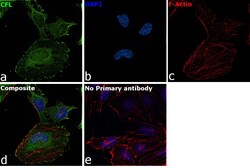
- Immunofluorescence analysis of Cofilin was performed using 70% confluent log phase HeLa cells. The cells were fixed with 4% paraformaldehyde for 10 minutes, permeabilized with 0.1% Triton™ X-100 for 15 minutes, and blocked with 2% BSA for 45 minutes at room temperature. The cells were labeled with Cofilin Polyclonal Antibody (Product # PA1-24931) at 1:1000 in 0.1% BSA, incubated at 4 degree celsius overnight and then labeled with Goat anti-Rabbit IgG (H+L) Superclonal™ Recombinant Secondary Antibody, Alexa Fluor® 488 conjugate (Product # A27034), (1:2000), for 45 minutes at room temperature (Panel a: Green). Nuclei (Panel b:Blue) were stained with ProLong™ Diamond Antifade Mountant with DAPI (Product # P36962). F-actin (Panel c: Red) was stained with Rhodamine Phalloidin (Product # R415, 1:300). Panel d represents the merged image showing cytoskeleton, plasma membrane, nucleus and cytoplasm localization. Panel e represents control cells with no primary antibody to assess background. The images were captured at 60X magnification.

Supportive validation
- Submitted by
- Invitrogen Antibodies (provider)
- Main image
- Experimental details
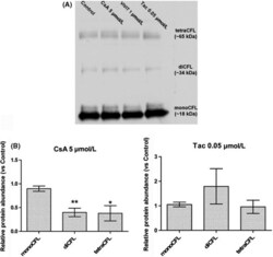
- Figure 3 Cyclosporine A (CsA), but not tacrolimus (Tac), induced changes in cofilin (CFL) concentration-to-actin. A, Western blot detection of formaldehyde-cross-linked CFL oligomers in Lilly Laboratories Porcine Kidney-1 lysates. B, Quantification of relative protein abundance of CFL oligomeric forms. Mean +- SEM. One-way ANOVA plus Dunnett's post-test (* P < .05, ** P < .01). Drug condition: 5 umol/L CsA, 0.05 umol/L Tac. Exposure time: 24 h (n = 5)
