Explore
 Validate
 Learn
Learn Western blot
Antibody data
- Antibody Data
- Antigen structure
- References [1]
- Comments [0]
- Validations
- Western blot [6]
- Immunocytochemistry [1]
- Immunohistochemistry [1]
Submit
Validation data
Reference
Comment
Report error
- Product number
- MA5-17275 - Provider product page

- Provider
- Invitrogen Antibodies
- Product name
- Cofilin Monoclonal Antibody (GT567)
- Antibody type
- Monoclonal
- Antigen
- Recombinant protein fragment
- Description
- Recommended positive controls: 293T, A431, HeLa, HepG2, A375, Neuro2A, C8D30, NIH-3T3, Raw264.7, C2C12, PC-12, Rat-2.
- Antibody clone number
- GT567
- Concentration
- 1 mg/mL
Submitted references ADF and cofilin-1 collaborate to promote cortical actin flow and the leader bleb-based migration of confined cells.
Ullo MF, Logue JS
eLife 2021 Jun 25;10
eLife 2021 Jun 25;10
No comments: Submit comment
Supportive validation
- Submitted by
- Invitrogen Antibodies (provider)
- Main image

- Experimental details
- Western blot analysis of Cofilin using A) 30 µg NIH-3T3 whole cell lysate (B) 30 µg JC whole cell lysate and C) 30 µg BCL-1 whole cell lysate. Samples were loaded onto a 12% SDS-PAGE gel and probed with a Cofilin monoclonal antibody (Product # MA5-17275) at a dilution of 1:1000.
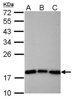
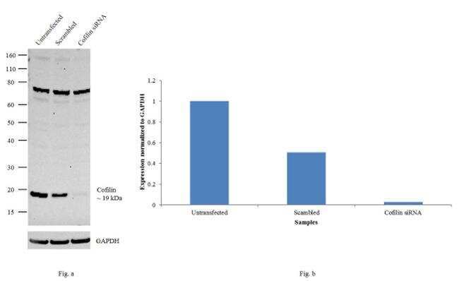
- Submitted by
- Invitrogen Antibodies (provider)
- Main image

- Experimental details
- Knockdown of cofilin was achieved by transfecting HeLa cells with cofilin specific siRNAs (Silencer® select Product # s2938, s2936). Western blot analysis (Fig. a) was performed using whole cell extracts from the Cofilin knockdown cells (lane 3), non-specific scrambled siRNA transfected cells (lane 2) and untransfected cells (lane 1). The blots were probed with cofilin monoclonal Antibody (Product # MA5-17275, 1:1000 dilution) and Goat anti-Mouse IgG (H+L) Superclonal™ Secondary Antibody, HRP conjugate (Product # A28177, 0.25 µg/ml 1:4000 dilution). Densitometric analysis of this western blot is shown in histogram (Fig. b). Decrease in signal upon siRNA mediated knock down confirms that antibody is specific to Cofilin.
- Submitted by
- Invitrogen Antibodies (provider)
- Main image
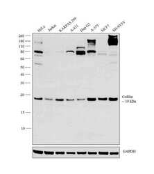
- Experimental details
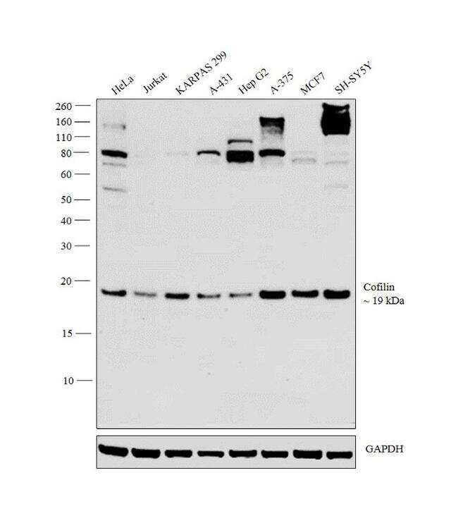
- Western blot analysis was performed on whole cell extracts (30 µg lysate) of HeLa (Lane 1), Jurkat (Lane 2), KARPASS 299 (Lane 3), A-431 (Lane 4), Hep G2 (Lane 5), A-375 (Lane 6), MCF7 (Lane 7) and SH-SY5Y (Lane 8). The blot was probed with Anti-Cofilin Monoclonal Antibody (Product # MA5-17275, 1:1000 dilution) and detected by chemiluminescence using Goat anti-Mouse IgG (H+L) Superclonal™ Secondary Antibody, HRP conjugate (Product # A28177, 0.25 µg/ml, 1:4000 dilution). A 19 kDa band corresponding to Cofilin with additional non specific bands at higher molecular weight were observed across the cell lines tested.
- Submitted by
- Invitrogen Antibodies (provider)
- Main image
- Experimental details
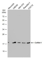
- Western Blot analysis of Cofilin was performed by separating 30 µg of various whole cell extracts by 12% SDS-PAGE. Proteins were transferred to a membrane and probed with a Cofilin Monoclonal Antibody (GT567) (Product # MA5-17275) at a dilution of 1:1000 and a HRP-conjugated anti-mouse IgG secondary antibody.

- Submitted by
- Invitrogen Antibodies (provider)
- Main image

- Experimental details
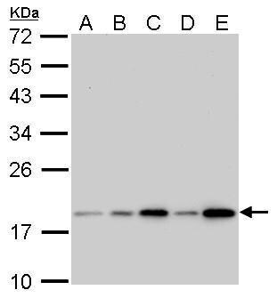
- Cofilin Monoclonal Antibody (GT567) detects CFL1 protein by western blot analysis. A. 30 µg 293T whole cell lysate/extract. B. 30 µg A431 whole cell lysate/extract. C. 30 µg HeLa whole cell lysate/extract. D. 30 µg HepG2 whole cell lysate/extract. E. 30 µg A375 whole cell lysate/extract.12% SDS-PAGE. Cofilin Monoclonal Antibody (GT567) (Product # MA5-17275) dilution: 1:1,000. The HRP-conjugated anti-mouse IgG antibody was used to detect the primary antibody.
- Submitted by
- Invitrogen Antibodies (provider)
- Main image
- Experimental details
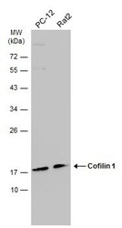
- Western Blot analysis of Cofilin was performed by separating 30 µg of various whole cell extracts by 12% SDS-PAGE. Proteins were transferred to a membrane and probed with a Cofilin Monoclonal Antibody (GT567) (Product # MA5-17275) at a dilution of 1:1000 and a HRP-conjugated anti-mouse IgG secondary antibody.

Supportive validation
- Submitted by
- Invitrogen Antibodies (provider)
- Main image
- Experimental details
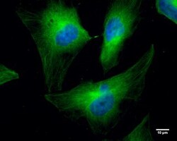
- Immunocytochemistry-Immunofluorescence analysis of Cofilin was performed in HeLa cells fixed in 4% paraformaldehyde at RT for 10 min. Green: Cofilin Monoclonal Antibody (GT567) (Product # MA5-17275) diluted at 1:300. Blue: Hoechst 33342 staining. Scale bar = 10 µm.

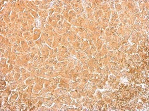
Supportive validation
- Submitted by
- Invitrogen Antibodies (provider)
- Main image
- Experimental details
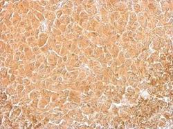
- Cofilin Monoclonal Antibody (GT567) detects CFL1 protein at cytosol on HBL435 xenograft by immunohistochemical analysis. Sample: Paraffin-embedded HBL435 xenograft. Cofilin Monoclonal Antibody (GT567) (Product # MA5-17275) dilution: 1:200. Antigen Retrieval: EDTA based buffer, pH 8.0, 15 min.