Explore
 Validate
 Learn
Learn Western blot
Antibody data
- Antibody Data
- Antigen structure
- References [4]
- Comments [0]
- Validations
- Western blot [2]
- ELISA [1]
- Immunocytochemistry [1]
- Immunohistochemistry [1]
Submit
Validation data
Reference
Comment
Report error
- Product number
- H00001072-M04 - Provider product page

- Provider
- Novus Biologicals
- Proper citation
- Novus Cat#H00001072-M04, RRID:AB_668974
- Product name
- Mouse Monoclonal Cofilin Antibody
- Antibody type
- Monoclonal
- Description
- IgG purified. cofilin 1 (non-muscle)
- Reactivity
- Human, Mouse
- Host
- Mouse
- Isotype
- IgG
- Vial size
- 0.1 mg
- Storage
- Aliquot and store at -20C or -80C. Avoid freeze-thaw cycles.
Submitted references Determination of urine cofilin-1 level in acute kidney injury using a high-throughput localized surface plasmon-coupled fluorescence biosensor.
Proteomic analysis of human epithelial lining fluid by microfluidics-based nanoLC-MS/MS: a feasibility study.
Identification of phosphorylated serine-15 and -82 residues of HSPB1 in 5-fluorouracil-resistant colorectal cancer cells by proteomics.
Overexpression of cofilin 1 can predict progression-free survival in patients with epithelial ovarian cancer receiving standard therapy.
Chang YF, Chao CH, Lin LY, Tsai CH, Chou C, Lee YJ
Journal of biomedical optics 2014 Jan;19(1):011004
Journal of biomedical optics 2014 Jan;19(1):011004
Proteomic analysis of human epithelial lining fluid by microfluidics-based nanoLC-MS/MS: a feasibility study.
Franciosi L, Govorukhina N, Fusetti F, Poolman B, Lodewijk ME, Timens W, Postma D, ten Hacken N, Bischoff R
Electrophoresis 2013 Sep;34(18):2683-94
Electrophoresis 2013 Sep;34(18):2683-94
Identification of phosphorylated serine-15 and -82 residues of HSPB1 in 5-fluorouracil-resistant colorectal cancer cells by proteomics.
Sakai A, Otani M, Miyamoto A, Yoshida H, Furuya E, Tanigawa N
Journal of proteomics 2012 Jan 4;75(3):806-18
Journal of proteomics 2012 Jan 4;75(3):806-18
Overexpression of cofilin 1 can predict progression-free survival in patients with epithelial ovarian cancer receiving standard therapy.
Nishimura S, Tsuda H, Kataoka F, Arao T, Nomura H, Chiyoda T, Susumu N, Nishio K, Aoki D
Human pathology 2011 Apr;42(4):516-21
Human pathology 2011 Apr;42(4):516-21
No comments: Submit comment
Supportive validation
- Submitted by
- Novus Biologicals (provider)
- Main image
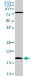
- Experimental details
- Western Blot: Cofilin Antibody (1A1) [H00001072-M04] - Analysis of CFL1 expression in NIH/3T3 (Cat # L018V1).

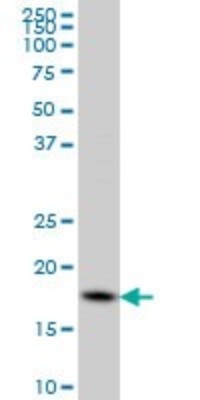
- Submitted by
- Novus Biologicals (provider)
- Main image

- Experimental details
- Western Blot: Cofilin Antibody (1A1) [H00001072-M04] - Analysis of CFL1 expression in HeLa (Cat # L013V1).
Supportive validation
- Submitted by
- Novus Biologicals (provider)
- Main image
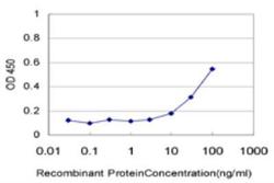
- Experimental details
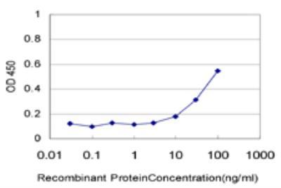
- ELISA: Cofilin Antibody (1A1) [H00001072-M04] - Detection limit for recombinant GST tagged CFL1 is approximately 10ng/ml as a capture antibody.
Supportive validation
- Submitted by
- Novus Biologicals (provider)
- Main image
- Experimental details
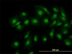
- Immunocytochemistry/Immunofluorescence: Cofilin Antibody (1A1) [H00001072-M04] - Analysis of monoclonal antibody to CFL1 on HeLa cell. Antibody concentration 10 ug/ml

Supportive validation
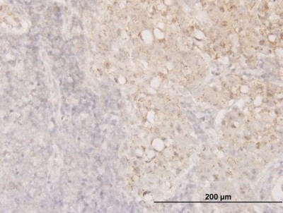
- Submitted by
- Novus Biologicals (provider)
- Main image

- Experimental details
- Immunohistochemistry-Paraffin: Cofilin Antibody (1A1) [H00001072-M04] - Analysis of monoclonal antibody to CFL1 on formalin-fixed paraffin-embedded human breast cancer. Antibody concentration 1.5 ug/ml