Explore
 Validate
 Learn
Learn Western blot
 Immunocytochemistry
Antibody data
- Antibody Data
- Antigen structure
- References [1]
- Comments [0]
- Validations
- Immunocytochemistry [5]
- Immunohistochemistry [1]
- Other assay [2]
Submit
Validation data
Reference
Comment
Report error
- Product number
- PA5-27627 - Provider product page

- Provider
- Invitrogen Antibodies
- Product name
- Cofilin Polyclonal Antibody
- Antibody type
- Polyclonal
- Antigen
- Recombinant full-length protein
- Description
- Recommended positive controls: 293T, A431, NIH-3T3, PC-12, Rat2. Predicted reactivity: Mouse (98%), Rat (99%), Pig (99%), Sheep (99%), Rhesus Monkey (99%), Bovine (99%). Store product as a concentrated solution. Centrifuge briefly prior to opening the vial.
- Reactivity
- Human, Mouse, Rat
- Host
- Rabbit
- Isotype
- IgG
- Vial size
- 100 μL
- Concentration
- 1 mg/mL
- Storage
- Store at 4°C short term. For long term storage, store at -20°C, avoiding freeze/thaw cycles.
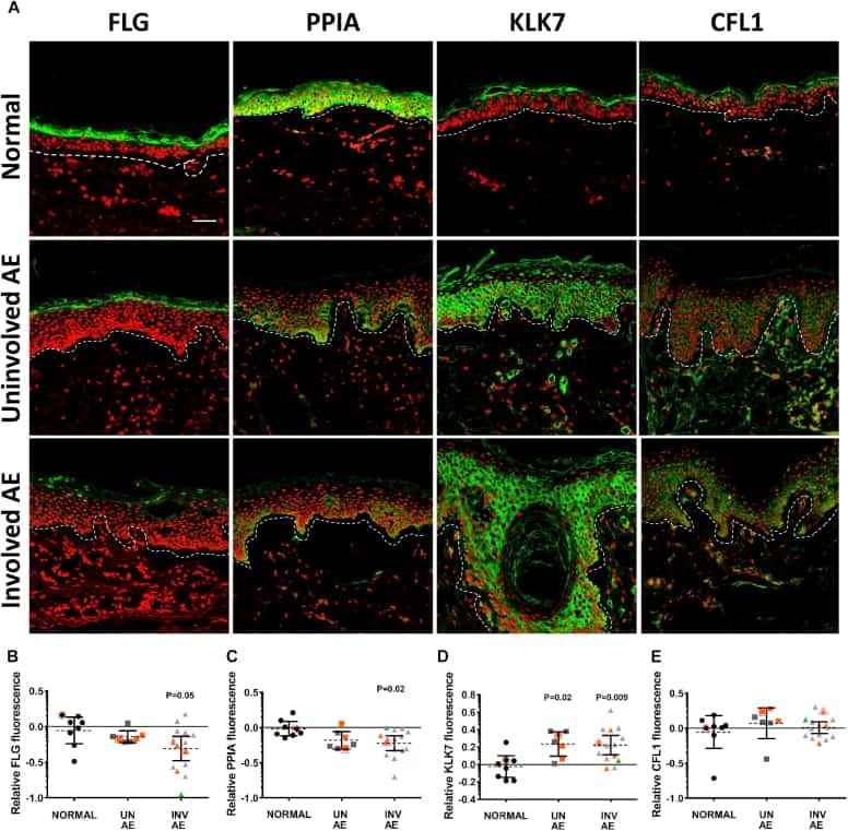
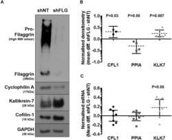
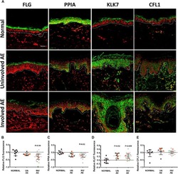
Submitted references Proteomic analysis of filaggrin deficiency identifies molecular signatures characteristic of atopic eczema.
Elias MS, Long HA, Newman CF, Wilson PA, West A, McGill PJ, Wu KC, Donaldson MJ, Reynolds NJ
The Journal of allergy and clinical immunology 2017 Nov;140(5):1299-1309
The Journal of allergy and clinical immunology 2017 Nov;140(5):1299-1309
No comments: Submit comment
Supportive validation
- Submitted by
- Invitrogen Antibodies (provider)
- Main image
- Experimental details
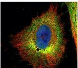
- Immunofluorescent analysis of Cofilin 1 (non-muscle) in paraformaldehyde-fixed HeLa cells using a Cofilin 1 (non-muscle) polyclonal antibody (Product # PA5-27627) (Green) at a 1:500 dilution. Alpha-tubulin filaments were labeled with Product # PA5-29281 (Red) at a 1:2000.
- Submitted by
- Invitrogen Antibodies (provider)
- Main image

- Experimental details
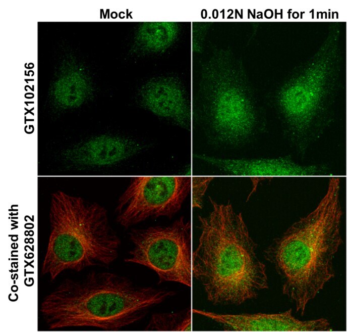
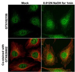
- Immunocytochemistry-Immunofluorescence analysis of Cofilin Cofilin in HeLa cells mock (left) and treated with 0.012 N NaOH/PBS for 1 min (right). Cells were fixed in 4% paraformaldehyde at RT for 15 min. Green: Cofilin Polyclonal Antibody (Product # PA5-27627) diluted at 1:1000. Red: alpha Tubulin, a cytoskeleton marker, stained by alpha Tubulin antibody.
- Submitted by
- Invitrogen Antibodies (provider)
- Main image

- Experimental details
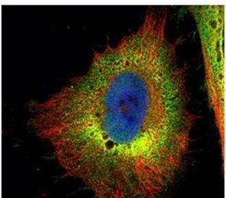
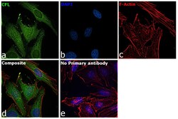
- Immunofluorescence analysis of Cofilin was performed using 70 confluent log phase HeLa cells. The cells were fixed with 4% paraformaldehyde for 10 minutes, permeabilized with 0.1% Triton™ X-100 for 15 minutes, and blocked with 2% BSA for 45 minutes at room temperature. The cells were labeled with Cofilin Polyclonal Antibody (Product # PA5-27627) at 1:100 in 0.1% BSA, incubated at 4 degree celsius overnight and then labeled with Goat anti-Rabbit IgG (H+L) Superclonal™ Recombinant Secondary Antibody, Alexa Fluor® 488 conjugate (Product # A27034), (1:2000), for 45 minutes at room temperature (Panel a: Green). Nuclei (Panel b:Blue) were stained with ProLong™ Diamond Antifade Mountant with DAPI (Product # P36962). F-actin (Panel c: Blue) was stained with Rhodamine Phalloidin (Product # R415, 1:300). Panel d represents the merged image showing cytoskeleton, plasma membrane, nucleus and cytoplasm localization. Panel e represents control cells with no primary antibody to assess background. The images were captured at 60 magnification.

- Submitted by
- Invitrogen Antibodies (provider)
- Main image
- Experimental details
- Immunocytochemistry-Immunofluorescence analysis of Cofilin Cofilin in HeLa cells mock (left) and treated with 0.012 N NaOH/PBS for 1 min (right). Cells were fixed in 4% paraformaldehyde at RT for 15 min. Green: Cofilin Polyclonal Antibody (Product # PA5-27627) diluted at 1:1000. Red: alpha Tubulin, a cytoskeleton marker, stained by alpha Tubulin antibody.

- Submitted by
- Invitrogen Antibodies (provider)
- Main image
- Experimental details
- Immunofluorescence analysis of Cofilin was performed using 70 confluent log phase HeLa cells. The cells were fixed with 4% paraformaldehyde for 10 minutes, permeabilized with 0.1% Triton™ X-100 for 15 minutes, and blocked with 2% BSA for 45 minutes at room temperature. The cells were labeled with Cofilin Polyclonal Antibody (Product # PA5-27627) at 1:100 in 0.1% BSA, incubated at 4 degree celsius overnight and then labeled with Goat anti-Rabbit IgG (Heavy Chain) Superclonal™ Recombinant Secondary Antibody, Alexa Fluor® 488 conjugate (Product # A27034), (1:2000), for 45 minutes at room temperature (Panel a: Green). Nuclei (Panel b:Blue) were stained with ProLong™ Diamond Antifade Mountant with DAPI (Product # P36962). F-actin (Panel c: Blue) was stained with Rhodamine Phalloidin (Product # R415, 1:300). Panel d represents the merged image showing cytoskeleton, plasma membrane, nucleus and cytoplasm localization. Panel e represents control cells with no primary antibody to assess background. The images were captured at 60 magnification.

Supportive validation
- Submitted by
- Invitrogen Antibodies (provider)
- Main image
- Experimental details
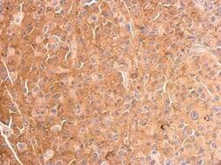
- Cofilin Polyclonal Antibody detects CFL1 protein at cytosol on HBL435 xenograft by immunohistochemical analysis. Sample: Paraffin-embedded HBL435 xenograft. Cofilin Polyclonal Antibody (Product # PA5-27627) dilution: 1:500. Antigen Retrieval: EDTA based buffer, pH 8.0, 15 min.

Supportive validation
- Submitted by
- Invitrogen Antibodies (provider)
- Main image
- Experimental details
- NULL

- Submitted by
- Invitrogen Antibodies (provider)
- Main image
- Experimental details
- NULL