Explore
 Validate
 Learn
Learn Western blot
Antibody data
- Antibody Data
- Antigen structure
- References [2]
- Comments [0]
- Validations
- Western blot [1]
- Immunocytochemistry [1]
- Immunohistochemistry [1]
Submit
Validation data
Reference
Comment
Report error
- Product number
- AP3122a - Provider product page

- Provider
- Abcepta
- Proper citation
- Abgent Cat#AP3122a, RRID:AB_637621
- Product name
- Phospho-HER4(Y1162) Antibody
- Antibody type
- Polyclonal
- Antigen
- Synthetic peptide
- Description
- Peptide Affinity Purified Rabbit Polyclonal Antibody (Pab)
- Reactivity
- Human
- Host
- Rabbit
- Isotype
- IgG
- Vial size
- 400 µl
- Concentration
- 0.5 mg/ml
- Storage
- Maintain refrigerated at 2-8°C for up to 6 months. For long term storage store at -20°C in small aliquots to prevent freeze-thaw cycles.
Submitted references Analysis of the tyrosine kinome in melanoma reveals recurrent mutations in ERBB4.
Choice of fixative is crucial to successful immunohistochemical detection of phosphoproteins in paraffin-embedded tumor tissues.
Prickett TD, Agrawal NS, Wei X, Yates KE, Lin JC, Wunderlich JR, Cronin JC, Cruz P, Rosenberg SA, Samuels Y
Nature genetics 2009 Oct;41(10):1127-32
Nature genetics 2009 Oct;41(10):1127-32
Choice of fixative is crucial to successful immunohistochemical detection of phosphoproteins in paraffin-embedded tumor tissues.
Burns JA, Li Y, Cheney CA, Ou Y, Franlin-Pfeifer LL, Kuklin N, Zhang ZQ
The journal of histochemistry and cytochemistry : official journal of the Histochemistry Society 2009 Mar;57(3):257-64
The journal of histochemistry and cytochemistry : official journal of the Histochemistry Society 2009 Mar;57(3):257-64
No comments: Submit comment
Supportive validation
- Submitted by
- Abcepta (provider)
- Main image
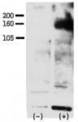
- Experimental details
- "Phospho-HER4-Y1162 Antibody (cat# AP3122a)was used to detect Phospho-HER4-Y1162 in FG pancreatic carcinoma cell line lysate. FG pancreatic carcinoma cells treated with or without EGF (50ng/ml) for 15 min Phospho-HER4-Y1162 Antibody (cat# AP3122a) was used at 1:750 in 3% BSA. Data and protocol kindly provided by Dr. Weis of Cheresh Lab, UCSD."
- Primary Ab dilution
- 1:1000

Supportive validation
- Submitted by
- Abcepta (provider)
- Main image
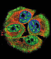
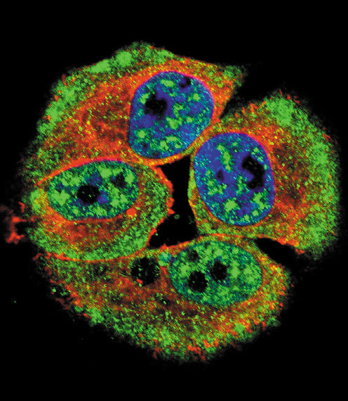
- Experimental details
- Confocal immunofluorescent analysis of Phospho-HER4-Y1162 Antibody(Cat#AP3122a) with MCF-7 cell followed by Alexa Fluor 488-conjugated goat anti-rabbit lgG (green). Actin filaments have been labeled with Alexa Fluor 555 phalloidin (red).DAPI was used to stain the cell nuclear (blue).
- Primary Ab dilution
- 1:10~50
Supportive validation
- Submitted by
- Abcepta (provider)
- Main image

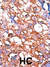
- Experimental details
- "Formalin-fixed and paraffin-embedded human cancer tissue reacted with the primary antibody, which was peroxidase-conjugated to the secondary antibody, followed by AEC staining. This data demonstrates the use of this antibody for immunohistochemistry; clinical relevance has not been evaluated. BC = breast carcinoma; HC = hepatocarcinoma."
- Primary Ab dilution
- 1:50~100