Explore
 Validate
 Learn
LearnMA1-861
antibody from Invitrogen Antibodies
Targeting: ERBB4
ALS19, HER4
 Western blot Immunocytochemistry
 Immunoprecipitation Immunohistochemistry Flow cytometry Other assay
Antibody data
- Antibody Data
- Antigen structure
- References [33]
- Comments [0]
- Validations
- Western blot [1]
- Immunocytochemistry [5]
- Immunohistochemistry [8]
- Flow cytometry [6]
- Other assay [1]
Submit
Validation data
Reference
Comment
Report error
- Product number
- MA1-861 - Provider product page

- Provider
- Invitrogen Antibodies
- Product name
- ErbB4 Monoclonal Antibody (HFR1)
- Antibody type
- Monoclonal
- Antigen
- Synthetic peptide
- Description
- MA1-861 detects Her-4 from human and mouse samples. MA1-861 has been successfully used in immunoprecipitation, immunohistochemical, and Western blotting procedures. By Western blot, this antibody detects ~185 kDa protein representing Her-4 from human A431 cell and mouse NIH3T3/HER4 cell extracts.This antibody has also been used in immunohistochemical, using paraffin embedded mouse NIH3T3 cells. MA1-861 immunizing peptide corresponds to amino acid residues 1250 to 1264 of human Her-4 oncoprotein.
- Reactivity
- Human, Mouse
- Host
- Mouse
- Isotype
- IgG
- Antibody clone number
- HFR1
- Vial size
- 200 μg
- Concentration
- 1 mg/mL
- Storage
- -20°C, Avoid Freeze/Thaw Cycles
Submitted references Screening for potential targets to reduce stenosis in bioprosthetic heart valves.
Nrg1β Released in Remote Ischemic Preconditioning Improves Myocardial Perfusion and Decreases Ischemia/Reperfusion Injury via ErbB2-Mediated Rescue of Endothelial Nitric Oxide Synthase and Abrogation of Trx2 Autophagy.
The reemergence of long-term potentiation in aged Alzheimer's disease mouse model.
Prognostic and biological significance of proliferation and HER2 expression in the luminal class of breast cancer.
Expression of the epidermal growth factor system in human middle ear cholesteatoma.
CYT-1 isoform of ErbB4 is an independent prognostic factor in serous ovarian cancer and selectively promotes ovarian cancer cell growth in vitro.
Cystatin M loss is associated with the losses of estrogen receptor, progesterone receptor, and HER4 in invasive breast cancer.
Overexpression of c-erbB2 is a negative prognostic factor in anaplastic astrocytomas.
HER-family gene amplification and expression in resected pancreatic cancer.
Expression of tumor necrosis factor alpha converting enzyme in endocrine cancers.
Distinctive expression pattern of ErbB family receptors signifies an aggressive variant of bladder cancer.
The HER4/4ICD estrogen receptor coactivator and BH3-only protein is an effector of tamoxifen-induced apoptosis.
Coexpression of c-erbB 1-4 receptor proteins in human glioblastomas. An immunohistochemical study.
Differential nuclear localization and kinase activity of alternative ErbB4 intracellular domains.
Phosphorylated HER-2 tyrosine kinase and Her-2/neu gene amplification as predictive factors of response to trastuzumab in patients with HER-2 overexpressing metastatic breast cancer (MBC).
Expression of the epidermal growth factor system in endometrioid endometrial cancer.
Essential erbB family phosphorylation in osteosarcoma as a target for CI-1033 inhibition.
Morphological and immunophenotypic analysis of breast carcinomas with basal and myoepithelial differentiation.
The role of HER1-HER4 and EGFRvIII in hormone-refractory prostate cancer.
Prognostic significance of HER3 and HER4 protein expression in colorectal adenocarcinomas.
Ovarian granulosa cell tumors frequently express EGFR (Her-1), Her-3, and Her-4: An immunohistochemical study.
High-throughput protein expression analysis using tissue microarray technology of a large well-characterised series identifies biologically distinct classes of breast cancer confirming recent cDNA expression analyses.
Correlation between human epidermal growth factor receptor family (EGFR, HER2, HER3, HER4), phosphorylated Akt (P-Akt), and clinical outcomes after radiation therapy in carcinoma of the cervix.
NRG1 gene rearrangements in clinical breast cancer: identification of an adjacent novel amplicon associated with poor prognosis.
Coexpression of the type 1 growth factor receptor family members HER-1, HER-2, and HER-3 has a synergistic negative prognostic effect on breast carcinoma survival.
Paclitaxel and carboplatin as first-line chemotherapy combined with gefitinib (IRESSA) in patients with advanced breast cancer: a phase I/II study conducted by the Hellenic Cooperative Oncology Group.
H-RAS V12-induced radioresistance in HCT116 colon carcinoma cells is heregulin dependent.
Expression of the epidermal growth factor system in human endometrium during the menstrual cycle.
Expression and co-expression of the members of the epidermal growth factor receptor (EGFR) family in invasive breast carcinoma.
Exquisite antitumour response to trastuzumab in a patient with no evidence of Ras-Raf-MAPK and PI3K-Akt pathways activation.
A recurrent chromosome translocation breakpoint in breast and pancreatic cancer cell lines targets the neuregulin/NRG1 gene.
CD44 anchors the assembly of matrilysin/MMP-7 with heparin-binding epidermal growth factor precursor and ErbB4 and regulates female reproductive organ remodeling.
Radiosensitization of malignant glioma cells through overexpression of dominant-negative epidermal growth factor receptor.
Foth R, Shomroni O, Sigler M, Hörer J, Cleuziou J, Paul T, Eildermann K
Scientific reports 2021 Jan 28;11(1):2464
Scientific reports 2021 Jan 28;11(1):2464
Nrg1β Released in Remote Ischemic Preconditioning Improves Myocardial Perfusion and Decreases Ischemia/Reperfusion Injury via ErbB2-Mediated Rescue of Endothelial Nitric Oxide Synthase and Abrogation of Trx2 Autophagy.
Kundumani-Sridharan V, Subramani J, Owens C, Das KC
Arteriosclerosis, thrombosis, and vascular biology 2021 Aug;41(8):2293-2314
Arteriosclerosis, thrombosis, and vascular biology 2021 Aug;41(8):2293-2314
The reemergence of long-term potentiation in aged Alzheimer's disease mouse model.
Huh S, Baek SJ, Lee KH, Whitcomb DJ, Jo J, Choi SM, Kim DH, Park MS, Lee KH, Kim BC
Scientific reports 2016 Jul 5;6:29152
Scientific reports 2016 Jul 5;6:29152
Prognostic and biological significance of proliferation and HER2 expression in the luminal class of breast cancer.
Jerjees DA, Alabdullah M, Green AR, Alshareeda A, Macmillan RD, Ellis IO, Rakha EA
Breast cancer research and treatment 2014 Jun;145(2):317-30
Breast cancer research and treatment 2014 Jun;145(2):317-30
Expression of the epidermal growth factor system in human middle ear cholesteatoma.
Thorup MB, Munk M, Poulsen SS, Gaihede M, Nexo E, Sorensen BS, Ovesen T
Acta oto-laryngologica 2014 Feb;134(2):124-34
Acta oto-laryngologica 2014 Feb;134(2):124-34
CYT-1 isoform of ErbB4 is an independent prognostic factor in serous ovarian cancer and selectively promotes ovarian cancer cell growth in vitro.
Paatero I, Lassus H, Junttila TT, Kaskinen M, Bützow R, Elenius K
Gynecologic oncology 2013 Apr;129(1):179-87
Gynecologic oncology 2013 Apr;129(1):179-87
Cystatin M loss is associated with the losses of estrogen receptor, progesterone receptor, and HER4 in invasive breast cancer.
Ko E, Park SE, Cho EY, Kim Y, Hwang JA, Lee YS, Nam SJ, Bang S, Park J, Kim DH
Breast cancer research : BCR 2010;12(6):R100
Breast cancer research : BCR 2010;12(6):R100
Overexpression of c-erbB2 is a negative prognostic factor in anaplastic astrocytomas.
Gulati S, Ytterhus B, Granli US, Gulati M, Lydersen S, Torp SH
Diagnostic pathology 2010 Mar 23;5:18
Diagnostic pathology 2010 Mar 23;5:18
HER-family gene amplification and expression in resected pancreatic cancer.
te Velde EA, Franke AC, van Hillegersberg R, Elshof SM, de Weger RW, Borel Rinkes IH, van Diest PJ
European journal of surgical oncology : the journal of the European Society of Surgical Oncology and the British Association of Surgical Oncology 2009 Oct;35(10):1098-104
European journal of surgical oncology : the journal of the European Society of Surgical Oncology and the British Association of Surgical Oncology 2009 Oct;35(10):1098-104
Expression of tumor necrosis factor alpha converting enzyme in endocrine cancers.
Kirkegaard T, Naresh A, Sabine VS, Tovey SM, Edwards J, Dunne B, Cooke TG, Jones FE, Bartlett JM
American journal of clinical pathology 2008 May;129(5):735-43
American journal of clinical pathology 2008 May;129(5):735-43
Distinctive expression pattern of ErbB family receptors signifies an aggressive variant of bladder cancer.
Kassouf W, Black PC, Tuziak T, Bondaruk J, Lee S, Brown GA, Adam L, Wei C, Baggerly K, Bar-Eli M, McConkey D, Czerniak B, Dinney CP
The Journal of urology 2008 Jan;179(1):353-8
The Journal of urology 2008 Jan;179(1):353-8
The HER4/4ICD estrogen receptor coactivator and BH3-only protein is an effector of tamoxifen-induced apoptosis.
Naresh A, Thor AD, Edgerton SM, Torkko KC, Kumar R, Jones FE
Cancer research 2008 Aug 1;68(15):6387-95
Cancer research 2008 Aug 1;68(15):6387-95
Coexpression of c-erbB 1-4 receptor proteins in human glioblastomas. An immunohistochemical study.
Torp SH, Gulati S, Johannessen E, Dalen A
Journal of experimental & clinical cancer research : CR 2007 Sep;26(3):353-9
Journal of experimental & clinical cancer research : CR 2007 Sep;26(3):353-9
Differential nuclear localization and kinase activity of alternative ErbB4 intracellular domains.
Sundvall M, Peri L, Määttä JA, Tvorogov D, Paatero I, Savisalo M, Silvennoinen O, Yarden Y, Elenius K
Oncogene 2007 Oct 18;26(48):6905-14
Oncogene 2007 Oct 18;26(48):6905-14
Phosphorylated HER-2 tyrosine kinase and Her-2/neu gene amplification as predictive factors of response to trastuzumab in patients with HER-2 overexpressing metastatic breast cancer (MBC).
Giuliani R, Durbecq V, Di Leo A, Paesmans M, Larsimont D, Leroy JY, Borms M, Vindevoghel A, Jerusalem G, D'Hondt V, Dirix L, Canon JL, Richard V, Cocquyt V, Majois F, Reginster M, Demol J, Kains JP, Delree P, Keppens C, Sotiriou C, Piccart MJ, Cardoso F
European journal of cancer (Oxford, England : 1990) 2007 Mar;43(4):725-35
European journal of cancer (Oxford, England : 1990) 2007 Mar;43(4):725-35
Expression of the epidermal growth factor system in endometrioid endometrial cancer.
Ejskjaer K, Sørensen BS, Poulsen SS, Forman A, Nexø E, Mogensen O
Gynecologic oncology 2007 Jan;104(1):158-67
Gynecologic oncology 2007 Jan;104(1):158-67
Essential erbB family phosphorylation in osteosarcoma as a target for CI-1033 inhibition.
Hughes DP, Thomas DG, Giordano TJ, McDonagh KT, Baker LH
Pediatric blood & cancer 2006 May 1;46(5):614-23
Pediatric blood & cancer 2006 May 1;46(5):614-23
Morphological and immunophenotypic analysis of breast carcinomas with basal and myoepithelial differentiation.
Rakha EA, Putti TC, Abd El-Rehim DM, Paish C, Green AR, Powe DG, Lee AH, Robertson JF, Ellis IO
The Journal of pathology 2006 Mar;208(4):495-506
The Journal of pathology 2006 Mar;208(4):495-506
The role of HER1-HER4 and EGFRvIII in hormone-refractory prostate cancer.
Edwards J, Traynor P, Munro AF, Pirret CF, Dunne B, Bartlett JM
Clinical cancer research : an official journal of the American Association for Cancer Research 2006 Jan 1;12(1):123-30
Clinical cancer research : an official journal of the American Association for Cancer Research 2006 Jan 1;12(1):123-30
Prognostic significance of HER3 and HER4 protein expression in colorectal adenocarcinomas.
Kountourakis P, Pavlakis K, Psyrri A, Rontogianni D, Xiros N, Patsouris E, Pectasides D, Economopoulos T
BMC cancer 2006 Feb 28;6:46
BMC cancer 2006 Feb 28;6:46
Ovarian granulosa cell tumors frequently express EGFR (Her-1), Her-3, and Her-4: An immunohistochemical study.
Leibl S, Bodo K, Gogg-Kammerer M, Hrzenjak A, Petru E, Winter R, Denk H, Moinfar F
Gynecologic oncology 2006 Apr;101(1):18-23
Gynecologic oncology 2006 Apr;101(1):18-23
High-throughput protein expression analysis using tissue microarray technology of a large well-characterised series identifies biologically distinct classes of breast cancer confirming recent cDNA expression analyses.
Abd El-Rehim DM, Ball G, Pinder SE, Rakha E, Paish C, Robertson JF, Macmillan D, Blamey RW, Ellis IO
International journal of cancer 2005 Sep 1;116(3):340-50
International journal of cancer 2005 Sep 1;116(3):340-50
Correlation between human epidermal growth factor receptor family (EGFR, HER2, HER3, HER4), phosphorylated Akt (P-Akt), and clinical outcomes after radiation therapy in carcinoma of the cervix.
Lee CM, Shrieve DC, Zempolich KA, Lee RJ, Hammond E, Handrahan DL, Gaffney DK
Gynecologic oncology 2005 Nov;99(2):415-21
Gynecologic oncology 2005 Nov;99(2):415-21
NRG1 gene rearrangements in clinical breast cancer: identification of an adjacent novel amplicon associated with poor prognosis.
Prentice LM, Shadeo A, Lestou VS, Miller MA, deLeeuw RJ, Makretsov N, Turbin D, Brown LA, Macpherson N, Yorida E, Cheang MC, Bentley J, Chia S, Nielsen TO, Gilks CB, Lam W, Huntsman DG
Oncogene 2005 Nov 10;24(49):7281-9
Oncogene 2005 Nov 10;24(49):7281-9
Coexpression of the type 1 growth factor receptor family members HER-1, HER-2, and HER-3 has a synergistic negative prognostic effect on breast carcinoma survival.
Wiseman SM, Makretsov N, Nielsen TO, Gilks B, Yorida E, Cheang M, Turbin D, Gelmon K, Huntsman DG
Cancer 2005 May 1;103(9):1770-7
Cancer 2005 May 1;103(9):1770-7
Paclitaxel and carboplatin as first-line chemotherapy combined with gefitinib (IRESSA) in patients with advanced breast cancer: a phase I/II study conducted by the Hellenic Cooperative Oncology Group.
Fountzilas G, Pectasides D, Kalogera-Fountzila A, Skarlos D, Kalofonos HP, Papadimitriou C, Bafaloukos D, Lambropoulos S, Papadopoulos S, Kourea H, Markopoulos C, Linardou H, Mavroudis D, Briasoulis E, Pavlidis N, Razis E, Kosmidis P, Gogas H
Breast cancer research and treatment 2005 Jul;92(1):1-9
Breast cancer research and treatment 2005 Jul;92(1):1-9
H-RAS V12-induced radioresistance in HCT116 colon carcinoma cells is heregulin dependent.
Carón RW, Yacoub A, Zhu X, Mitchell C, Han SI, Sasazuki T, Shirasawa S, Hagan MP, Grant S, Dent P
Molecular cancer therapeutics 2005 Feb;4(2):243-55
Molecular cancer therapeutics 2005 Feb;4(2):243-55
Expression of the epidermal growth factor system in human endometrium during the menstrual cycle.
Ejskjaer K, Sørensen BS, Poulsen SS, Mogensen O, Forman A, Nexø E
Molecular human reproduction 2005 Aug;11(8):543-51
Molecular human reproduction 2005 Aug;11(8):543-51
Expression and co-expression of the members of the epidermal growth factor receptor (EGFR) family in invasive breast carcinoma.
Abd El-Rehim DM, Pinder SE, Paish CE, Bell JA, Rampaul RS, Blamey RW, Robertson JF, Nicholson RI, Ellis IO
British journal of cancer 2004 Oct 18;91(8):1532-42
British journal of cancer 2004 Oct 18;91(8):1532-42
Exquisite antitumour response to trastuzumab in a patient with no evidence of Ras-Raf-MAPK and PI3K-Akt pathways activation.
Mano MS, Awada A, Minisini A, Durbecq V, Di Leo A, Piccart M
Breast (Edinburgh, Scotland) 2004 Aug;13(4):347-9
Breast (Edinburgh, Scotland) 2004 Aug;13(4):347-9
A recurrent chromosome translocation breakpoint in breast and pancreatic cancer cell lines targets the neuregulin/NRG1 gene.
Adélaïde J, Huang HE, Murati A, Alsop AE, Orsetti B, Mozziconacci MJ, Popovici C, Ginestier C, Letessier A, Basset C, Courtay-Cahen C, Jacquemier J, Theillet C, Birnbaum D, Edwards PA, Chaffanet M
Genes, chromosomes & cancer 2003 Aug;37(4):333-45
Genes, chromosomes & cancer 2003 Aug;37(4):333-45
CD44 anchors the assembly of matrilysin/MMP-7 with heparin-binding epidermal growth factor precursor and ErbB4 and regulates female reproductive organ remodeling.
Yu WH, Woessner JF Jr, McNeish JD, Stamenkovic I
Genes & development 2002 Feb 1;16(3):307-23
Genes & development 2002 Feb 1;16(3):307-23
Radiosensitization of malignant glioma cells through overexpression of dominant-negative epidermal growth factor receptor.
Lammering G, Valerie K, Lin PS, Mikkelsen RB, Contessa JN, Feden JP, Farnsworth J, Dent P, Schmidt-Ullrich RK
Clinical cancer research : an official journal of the American Association for Cancer Research 2001 Mar;7(3):682-90
Clinical cancer research : an official journal of the American Association for Cancer Research 2001 Mar;7(3):682-90
No comments: Submit comment
Supportive validation
- Submitted by
- Invitrogen Antibodies (provider)
- Main image

- Experimental details
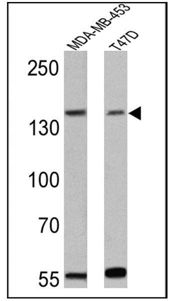
- Western blot analysis of ErbB4 was performed by loading 25 µg of MDA-MB-453 (lane 1) and T47D (lane 2) cell lysates onto an SDS polyacrylamide gel. Proteins were transferred to a PVDF membrane and blocked at 4ºC overnight. The membrane was probed with an ErbB4 monoclonal antibody (Product # MA1-861) at a dilution of 1:300 overnight at 4°C, washed in TBST, and probed with an HRP-conjugated goat anti-mouse IgG + IgM (H+L) cross-adsorbed secondary antibody for 1 hr at room temperature in the dark. Chemiluminescent detection was performed using Pierce ECL Plus Western Blotting Substrate (Product # 32132). Results show a band at ~185 kDa.
Supportive validation
- Submitted by
- Invitrogen Antibodies (provider)
- Main image
- Experimental details
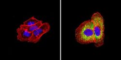
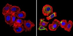
- Immunofluorescent analysis of ErbB4 (green) showing staining in the cytoplasm of A431 cells (right) compared to a negative control without primary antibody (left). Formalin-fixed cells were permeabilized with 0.1% Triton X-100 in TBS for 5-10 minutes and blocked with 3% BSA-PBS for 30 minutes at room temperature. Cells were probed with an ErbB4 monoclonal antibody (Product # MA1-861) in 3% BSA-PBS at a dilution of 1:50 and incubated overnight at 4ºC in a humidified chamber. Cells were washed with PBST and incubated with a DyLight-conjugated secondary antibody in PBS at room temperature in the dark. Actin was stained using Alexa Fluor 554 (red) and nuclei were stained with Hoechst or DAPI (blue). Images were taken at a magnification of 60x.

- Submitted by
- Invitrogen Antibodies (provider)
- Main image

- Experimental details
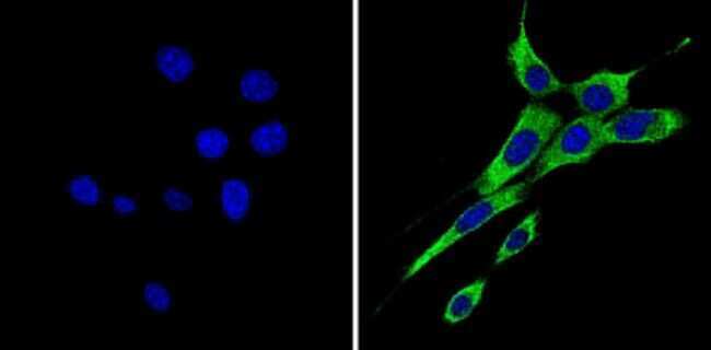
- Immunofluorescent analysis of ErbB4 (green) showing staining in the cytoplasm of NIH-3T3 cells (right) compared to a negative control without primary antibody (left). Formalin-fixed cells were permeabilized with 0.1% Triton X-100 in TBS for 5-10 minutes and blocked with 3% BSA-PBS for 30 minutes at room temperature. Cells were probed with an ErbB4 monoclonal antibody (Product # MA1-861) in 3% BSA-PBS at a dilution of 1:50 and incubated overnight at 4ºC in a humidified chamber. Cells were washed with PBST and incubated with a DyLight-conjugated secondary antibody in PBS at room temperature in the dark. Nuclei were stained with Hoechst or DAPI (blue). Images were taken at a magnification of 60x.
- Submitted by
- Invitrogen Antibodies (provider)
- Main image
- Experimental details
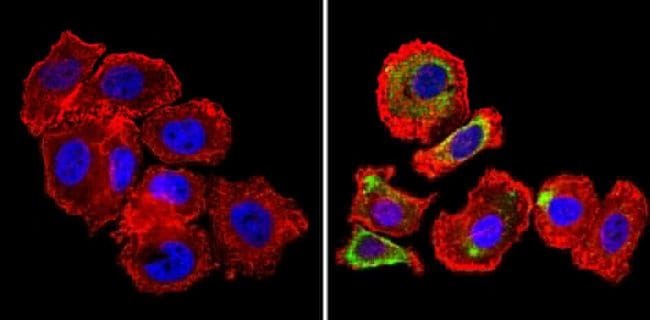
- Immunofluorescent analysis of ErbB4 (green) showing staining in the cytoplasm of SK-BR-3 cells (right) compared to a negative control without primary antibody (left). Formalin-fixed cells were permeabilized with 0.1% Triton X-100 in TBS for 5-10 minutes and blocked with 3% BSA-PBS for 30 minutes at room temperature. Cells were probed with an ErbB4 monoclonal antibody (Product # MA1-861) in 3% BSA-PBS at a dilution of 1:50 and incubated overnight at 4ºC in a humidified chamber. Cells were washed with PBST and incubated with a DyLight-conjugated secondary antibody in PBS at room temperature in the dark. Actin was stained using Alexa Fluor 554 (red) and nuclei were stained with Hoechst or DAPI (blue). Images were taken at a magnification of 60x.
- Submitted by
- Invitrogen Antibodies (provider)
- Main image

- Experimental details
- Immunofluorescent analysis of ErbB4 (green) showing staining in the cytoplasm of NIH-3T3 cells (right) compared to a negative control without primary antibody (left). Formalin-fixed cells were permeabilized with 0.1% Triton X-100 in TBS for 5-10 minutes and blocked with 3% BSA-PBS for 30 minutes at room temperature. Cells were probed with an ErbB4 monoclonal antibody (Product # MA1-861) in 3% BSA-PBS at a dilution of 1:50 and incubated overnight at 4ºC in a humidified chamber. Cells were washed with PBST and incubated with a DyLight-conjugated secondary antibody in PBS at room temperature in the dark. Nuclei were stained with Hoechst or DAPI (blue). Images were taken at a magnification of 60x.

- Submitted by
- Invitrogen Antibodies (provider)
- Main image

- Experimental details
- Immunofluorescent analysis of ErbB4 (green) showing staining in the cytoplasm of SK-BR-3 cells (right) compared to a negative control without primary antibody (left). Formalin-fixed cells were permeabilized with 0.1% Triton X-100 in TBS for 5-10 minutes and blocked with 3% BSA-PBS for 30 minutes at room temperature. Cells were probed with an ErbB4 monoclonal antibody (Product # MA1-861) in 3% BSA-PBS at a dilution of 1:50 and incubated overnight at 4ºC in a humidified chamber. Cells were washed with PBST and incubated with a DyLight-conjugated secondary antibody in PBS at room temperature in the dark. Actin was stained using Alexa Fluor 554 (red) and nuclei were stained with Hoechst or DAPI (blue). Images were taken at a magnification of 60x.

Supportive validation
- Submitted by
- Invitrogen Antibodies (provider)
- Main image
- Experimental details
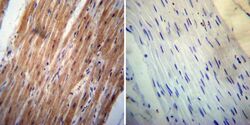
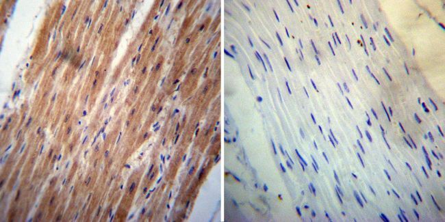
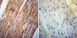
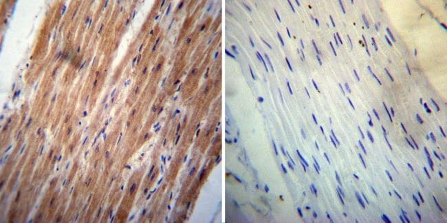
- Immunohistochemistry was performed on normal deparaffinized human Heart tissue. To expose target proteins, heat induced antigen retrieval was performed using 10mM sodium citrate (pH6.0) buffer, microwaved for 8-15 minutes. Following antigen retrieval tissues were blocked in 3% BSA-PBS for 30 minutes at room temperature. Tissues were then probed at a dilution of 1:200 with a mouse monoclonal antibody recognizing ErbB4 (Product # MA1-861) or without primary antibody (negative control) overnight at 4°C in a humidified chamber. Tissues were washed extensively with PBST and endogenous peroxidase activity was quenched with a peroxidase suppressor. Detection was performed using a biotin-conjugated secondary antibody and SA-HRP, followed by colorimetric detection using DAB. Tissues were counterstained with hematoxylin and prepped for mounting.
- Submitted by
- Invitrogen Antibodies (provider)
- Main image
- Experimental details
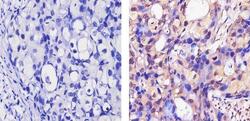
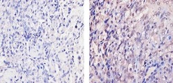
- Immunohistochemistry was performed on normal deparaffinized human Pancreas tissue. To expose target proteins, heat induced antigen retrieval was performed using 10mM sodium citrate (pH6.0) buffer, microwaved for 8-15 minutes. Following antigen retrieval tissues were blocked in 3% BSA-PBS for 30 minutes at room temperature. Tissues were then probed at a dilution of 1:200 with a mouse monoclonal antibody recognizing ErbB4 (Product # MA1-861) or without primary antibody (negative control) overnight at 4°C in a humidified chamber. Tissues were washed extensively with PBST and endogenous peroxidase activity was quenched with a peroxidase suppressor. Detection was performed using a biotin-conjugated secondary antibody and SA-HRP, followed by colorimetric detection using DAB. Tissues were counterstained with hematoxylin and prepped for mounting.

- Submitted by
- Invitrogen Antibodies (provider)
- Main image
- Experimental details
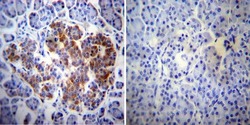
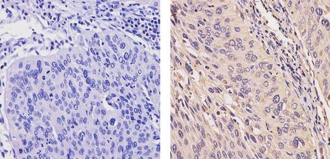
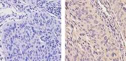
- Immunohistochemistry was performed on cancer biopsies of deparaffinized human Breast carcinoma tissue. To expose target proteins, heat induced antigen retrieval was performed using 10mM sodium citrate (pH6.0) buffer, microwaved for 8-15 minutes. Following antigen retrieval tissues were blocked in 3% BSA-PBS for 30 minutes at room temperature. Tissues were then probed at a dilution of 1:20 with a mouse monoclonal antibody recognizing ErbB4 (Product # MA1-861) or without primary antibody (negative control) overnight at 4°C in a humidified chamber. Tissues were washed extensively with PBST and endogenous peroxidase activity was quenched with a peroxidase suppressor. Detection was performed using a biotin-conjugated secondary antibody and SA-HRP, followed by colorimetric detection using DAB. Tissues were counterstained with hematoxylin and prepped for mounting.

- Submitted by
- Invitrogen Antibodies (provider)
- Main image
- Experimental details
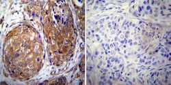
- Immunohistochemistry analysis of ErbB4 showing staining in the cytoplasm and nucleus of paraffin-embedded human breast carcinoma (right) compared to a negative control without primary antibody (left). To expose target proteins, antigen retrieval was performed using 10mM sodium citrate (pH 6.0), microwaved for 8-15 min. Following antigen retrieval, tissues were blocked in 3% H2O2-methanol for 15 min at room temperature, washed with ddH2O and PBS, and then probed with a ErbB4 Mouse Monoclonal Antibody (Product # MA1-861) diluted in 3% BSA-PBS at a dilution of 1:100 for 1 hour at 37ºC in a humidified chamber. Tissues were washed extensively in PBST and detection was performed using an HRP-conjugated secondary antibody followed by colorimetric detection using a DAB kit. Tissues were counterstained with hematoxylin and dehydrated with ethanol and xylene to prep for mounting.

- Submitted by
- Invitrogen Antibodies (provider)
- Main image
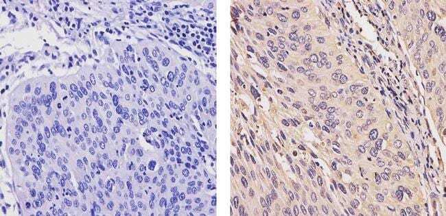
- Experimental details
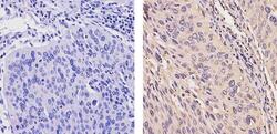
- Immunohistochemistry analysis of ErbB4 showing staining in the cytoplasm and nucleus of paraffin-embedded human cervix carcinoma (right) compared to a negative control without primary antibody (left). To expose target proteins, antigen retrieval was performed using 10mM sodium citrate (pH 6.0), microwaved for 8-15 min. Following antigen retrieval, tissues were blocked in 3% H2O2-methanol for 15 min at room temperature, washed with ddH2O and PBS, and then probed with a ErbB4 Mouse Monoclonal Antibody (Product # MA1-861) diluted in 3% BSA-PBS at a dilution of 1:100 for 1 hour at 37ºC in a humidified chamber. Tissues were washed extensively in PBST and detection was performed using an HRP-conjugated secondary antibody followed by colorimetric detection using a DAB kit. Tissues were counterstained with hematoxylin and dehydrated with ethanol and xylene to prep for mounting.
- Submitted by
- Invitrogen Antibodies (provider)
- Main image
- Experimental details
- Immunohistochemistry analysis of ErbB4 showing staining in the cytoplasm and nucleus of paraffin-embedded mouse placenta tissue (right) compared to a negative control without primary antibody (left). To expose target proteins, antigen retrieval was performed using 10mM sodium citrate (pH 6.0), microwaved for 8-15 min. Following antigen retrieval, tissues were blocked in 3% H2O2-methanol for 15 min at room temperature, washed with ddH2O and PBS, and then probed with a ErbB4 Mouse Monoclonal Antibody (Product # MA1-861) diluted in 3% BSA-PBS at a dilution of 1:100 for 1 hour at 37ºC in a humidified chamber. Tissues were washed extensively in PBST and detection was performed using an HRP-conjugated secondary antibody followed by colorimetric detection using a DAB kit. Tissues were counterstained with hematoxylin and dehydrated with ethanol and xylene to prep for mounting.

- Submitted by
- Invitrogen Antibodies (provider)
- Main image
- Experimental details
- Immunohistochemistry was performed on normal deparaffinized human Heart tissue. To expose target proteins, heat induced antigen retrieval was performed using 10mM sodium citrate (pH6.0) buffer, microwaved for 8-15 minutes. Following antigen retrieval tissues were blocked in 3% BSA-PBS for 30 minutes at room temperature. Tissues were then probed at a dilution of 1:200 with a mouse monoclonal antibody recognizing ErbB4 (Product # MA1-861) or without primary antibody (negative control) overnight at 4°C in a humidified chamber. Tissues were washed extensively with PBST and endogenous peroxidase activity was quenched with a peroxidase suppressor. Detection was performed using a biotin-conjugated secondary antibody and SA-HRP, followed by colorimetric detection using DAB. Tissues were counterstained with hematoxylin and prepped for mounting.
- Submitted by
- Invitrogen Antibodies (provider)
- Main image
- Experimental details
- Immunohistochemistry analysis of ErbB4 showing staining in the cytoplasm and nucleus of paraffin-embedded human cervix carcinoma (right) compared to a negative control without primary antibody (left). To expose target proteins, antigen retrieval was performed using 10mM sodium citrate (pH 6.0), microwaved for 8-15 min. Following antigen retrieval, tissues were blocked in 3% H2O2-methanol for 15 min at room temperature, washed with ddH2O and PBS, and then probed with a ErbB4 Mouse Monoclonal Antibody (Product # MA1-861) diluted in 3% BSA-PBS at a dilution of 1:100 for 1 hour at 37ºC in a humidified chamber. Tissues were washed extensively in PBST and detection was performed using an HRP-conjugated secondary antibody followed by colorimetric detection using a DAB kit. Tissues were counterstained with hematoxylin and dehydrated with ethanol and xylene to prep for mounting.
Supportive validation
- Submitted by
- Invitrogen Antibodies (provider)
- Main image
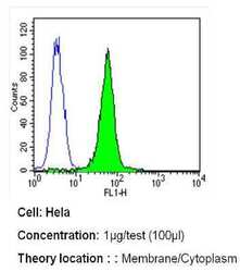
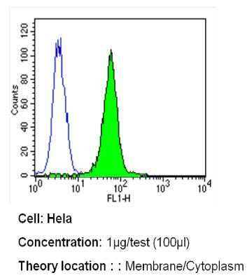
- Experimental details
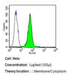
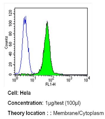
- Flow cytometry analysis of ErbB4 in Hela cells (green) compared to an isotype control (blue). Cells were harvested, adjusted to a concentration of 1-5x10^6 cells/mL, fixed with 2% paraformaldehyde and washed with PBS. Cells were blocked with a 2% solution of BSA-PBS for 30 min at room temperature and incubated with an ErbB4 monoclonal antibody (Product # MA1-861) at a dilution of 1 µg/test for 40 min at room temperature. Cells were then incubated for 40 min at room temperature in the dark using a Dylight 488-conjugated secondary antibody and re-suspended in PBS for FACS analysis.
- Submitted by
- Invitrogen Antibodies (provider)
- Main image

- Experimental details
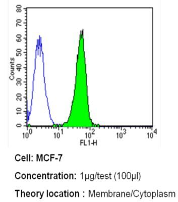
- Flow cytometry analysis of ErbB4 in MCF-7 cells (green) compared to an isotype control (blue). Cells were harvested, adjusted to a concentration of 1-5x10^6 cells/mL, fixed with 2% paraformaldehyde and washed with PBS. Cells were blocked with a 2% solution of BSA-PBS for 30 min at room temperature and incubated with an ErbB4 monoclonal antibody (Product # MA1-861) at a dilution of 1 µg/test for 40 min at room temperature. Cells were then incubated for 40 min at room temperature in the dark using a Dylight 488-conjugated secondary antibody and re-suspended in PBS for FACS analysis.

- Submitted by
- Invitrogen Antibodies (provider)
- Main image
- Experimental details
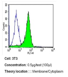
- Flow cytometry analysis of ErbB4 in NIH-3T3 cells (green) compared to an isotype control (blue). Cells were harvested, adjusted to a concentration of 1-5x10^6 cells/mL, fixed with 2% paraformaldehyde and washed with PBS. Cells were blocked with a 2% solution of BSA-PBS for 30 min at room temperature and incubated with an ErbB4 monoclonal antibody (Product # MA1-861) at a dilution of 0.5 µg/test for 40 min at room temperature. Cells were then incubated for 40 min at room temperature in the dark using a Dylight 488-conjugated secondary antibody and re-suspended in PBS for FACS analysis.

- Submitted by
- Invitrogen Antibodies (provider)
- Main image

- Experimental details
- Flow cytometry analysis of ErbB4 in MCF-7 cells (green) compared to an isotype control (blue). Cells were harvested, adjusted to a concentration of 1-5x10^6 cells/mL, fixed with 2% paraformaldehyde and washed with PBS. Cells were blocked with a 2% solution of BSA-PBS for 30 min at room temperature and incubated with an ErbB4 monoclonal antibody (Product # MA1-861) at a dilution of 1 µg/test for 40 min at room temperature. Cells were then incubated for 40 min at room temperature in the dark using a Dylight 488-conjugated secondary antibody and re-suspended in PBS for FACS analysis.
- Submitted by
- Invitrogen Antibodies (provider)
- Main image
- Experimental details
- Flow cytometry analysis of ErbB4 in Hela cells (green) compared to an isotype control (blue). Cells were harvested, adjusted to a concentration of 1-5x10^6 cells/mL, fixed with 2% paraformaldehyde and washed with PBS. Cells were blocked with a 2% solution of BSA-PBS for 30 min at room temperature and incubated with an ErbB4 monoclonal antibody (Product # MA1-861) at a dilution of 1 µg/test for 40 min at room temperature. Cells were then incubated for 40 min at room temperature in the dark using a Dylight 488-conjugated secondary antibody and re-suspended in PBS for FACS analysis.
Supportive validation
- Submitted by
- Invitrogen Antibodies (provider)
- Main image

- Experimental details
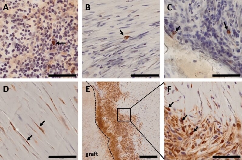
- Figure 3 Immunohistochemical staining of candidate genes (brown). Nuclei were stained with hematoxylin and appear in blue. All pictures show neotissue in the thickened area of the conduit wall. (A) Black arrow points to EGFR positive macrophage in granular tissue. (B) Black arrow points to ErbB4 positive macrophage in transition area between granular and fibrous tissue. (C) Black arrows point to ErbB4 positive macrophages in granular tissue. (D) Black arrows point to FLT4 positive, spindle shaped fibroblasts in fibrous tissue. (E) Overview picture of FLT4 staining. Graft material is located on the left side of the picture. A dotted line marks the border between graft and neo tissue. Neo tissue close to the graft was mostly granular, becoming more fibrous towards the lumen (right side in the picture). Here, granular tissue was generally intensively stained, whereas in fibrous tissue, staining was restricted to the fibroblasts. Square indicates the area that is zoomed in Fig. 10.1038/s41598-021-81340-2F. (F) Close up of square in Fig. 10.1038/s41598-021-81340-2E. Black arrows point to more intensively stained FLT4 positive macrophages. Scale bars: 50 um; scale bar Figure E: 200 um. Stainings were perfomed on n = 11 explanted conduits.