 Explore
 Validate
 Learn
Learn Western blot
 Immunocytochemistry
Antibody data
- Antibody Data
- Antigen structure
- References [2]
- Comments [0]
- Validations
- Immunocytochemistry [1]
Submit
Validation data
Reference
Comment
Report error
- Product number
- AF1131 - Provider product page

- Provider
- R&D Systems
- Product name
- Human ErbB4/Her4 Antibody
- Antibody type
- Polyclonal
- Description
- Antigen Affinity-purified. Detects human ErbB4/Her4 in direct ELISAs and Western blots. In direct ELISAs, approximately 25% cross-reactivity with recombinant mouse ErbB4 is observed, and less than 1% cross-reactivity with recombinant human (rh) ErbB2, rhErbB3, and rhEGF R is observed.
- Reactivity
- Human
- Host
- Goat
- Conjugate
- Unconjugated
- Antigen sequence
Q15303- Isotype
- IgG
- Vial size
- 100 ug
- Concentration
- LYOPH
- Storage
- Use a manual defrost freezer and avoid repeated freeze-thaw cycles. 12 months from date of receipt, -20 to -70 °C as supplied. 1 month, 2 to 8 °C under sterile conditions after reconstitution. 6 months, -20 to -70 °C under sterile conditions after reconstitution.
Submitted references Neuregulin-1-beta1 enters brain and spinal cord by receptor-mediated transport.
Neuregulin-1-beta1 enters brain and spinal cord by receptor-mediated transport.
Kastin AJ, Akerstrom V, Pan W
Journal of neurochemistry 2004 Feb;88(4):965-70
Journal of neurochemistry 2004 Feb;88(4):965-70
Neuregulin-1-beta1 enters brain and spinal cord by receptor-mediated transport.
Kastin AJ, Akerstrom V, Pan W
Journal of neurochemistry 2004 Feb;88(4):965-70
Journal of neurochemistry 2004 Feb;88(4):965-70
No comments: Submit comment
Supportive validation
- Submitted by
- R&D Systems (provider)
- Main image
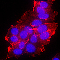
- Experimental details
- ErbB4/Her4 in MCF-7 Human Cell Line. ErbB4/Her4 was detected in immersion fixed MCF-7 human breast cancer cell line using Goat Anti-Human ErbB4/Her4 Antigen Affinity-purified Polyclonal Antibody (Catalog # AF1131) at 10 µg/mL for 3 hours at room temperature. Cells were stained using the NorthernLights™ 557-conjugated Anti-Goat IgG Secondary Antibody (red; Catalog # NL001) and counterstained with DAPI (blue). Specific staining was localized to plasma membrane. View our protocol for Fluorescent ICC Staining of Cells on Coverslips.
