Explore
 Validate
 Learn
Learn Western blot
 Immunocytochemistry
Antibody data
- Antibody Data
- Antigen structure
- References [3]
- Comments [0]
- Validations
- Immunocytochemistry [1]
- Immunohistochemistry [1]
Submit
Validation data
Reference
Comment
Report error
- Product number
- MAB1131 - Provider product page

- Provider
- R&D Systems
- Product name
- Human ErbB4/Her4 Antibody
- Antibody type
- Monoclonal
- Description
- Protein A or G purified from hybridoma culture supernatant. Detects human ErbB4/Her4 in direct ELISAs and Western blots. In direct ELISAs and Western blots, no cross-reactivity with recombinant human (rh) EGF R, rhErbB2, and rhErbB3 is observed.
- Reactivity
- Human
- Host
- Mouse
- Conjugate
- Unconjugated
- Antigen sequence
Q15303- Isotype
- IgG
- Antibody clone number
- 182803
- Vial size
- 500 ug
- Concentration
- LYOPH
- Storage
- Use a manual defrost freezer and avoid repeated freeze-thaw cycles. 12 months from date of receipt, -20 to -70 °C as supplied. 1 month, 2 to 8 °C under sterile conditions after reconstitution. 6 months, -20 to -70 °C under sterile conditions after reconstitution.
Submitted references miR-143, miR-222, and miR-452 are useful as tumor stratification and noninvasive diagnostic biomarkers for bladder cancer.
The fibroblast-derived paracrine factor neuregulin-1 has a novel role in regulating the constitutive color and melanocyte function in human skin.
Microbead arrays for the analysis of ErbB receptor tyrosine kinase activation and dimerization in breast cancer cells.
Puerta-Gil P, García-Baquero R, Jia AY, Ocaña S, Alvarez-Múgica M, Alvarez-Ossorio JL, Cordon-Cardo C, Cava F, Sánchez-Carbayo M
The American journal of pathology 2012 May;180(5):1808-15
The American journal of pathology 2012 May;180(5):1808-15
The fibroblast-derived paracrine factor neuregulin-1 has a novel role in regulating the constitutive color and melanocyte function in human skin.
Choi W, Wolber R, Gerwat W, Mann T, Batzer J, Smuda C, Liu H, Kolbe L, Hearing VJ
Journal of cell science 2010 Sep 15;123(Pt 18):3102-11
Journal of cell science 2010 Sep 15;123(Pt 18):3102-11
Microbead arrays for the analysis of ErbB receptor tyrosine kinase activation and dimerization in breast cancer cells.
Khan IH, Zhao J, Ghosh P, Ziman M, Sweeney C, Kung HJ, Luciw PA
Assay and drug development technologies 2010 Feb;8(1):27-36
Assay and drug development technologies 2010 Feb;8(1):27-36
No comments: Submit comment
Supportive validation
- Submitted by
- R&D Systems (provider)
- Main image
- Experimental details
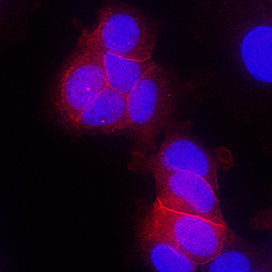
- ErbB4/Her4 in MCF-7 Human Cell Line. ErbB4/Her4 was detected in immersion fixed MCF-7 human breast cancer cell line using Mouse Anti-Human ErbB4/Her4 Monoclonal Antibody (Catalog # MAB1131) at 10 µg/mL for 3 hours at room temperature. Cells were stained using the NorthernLights™ 557-conjugated Anti-Mouse IgG Secondary Antibody (red; Catalog # NL007) and counterstained with DAPI (blue). Specific staining was localized to plasma membrane. View our protocol for Fluorescent ICC Staining of Cells on Coverslips.

Supportive validation
- Submitted by
- R&D Systems (provider)
- Main image
- Experimental details
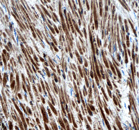
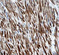
- ErbB4/Her4 in Human Colon Cancer Tissue. ErbB4/Her4 was detected in immersion fixed paraffin-embedded sections of human colon cancer tissue using Mouse Anti-Human ErbB4/Her4 Monoclonal Antibody (Catalog # MAB1131) at 15 µg/mL overnight at 4 °C. Tissue was stained using the Anti-Mouse HRP-DAB Cell & Tissue Staining Kit (brown; Catalog # CTS002) and counterstained with hematoxylin (blue). Specific staining was localized to smooth muscle cells. View our protocol for Chromogenic IHC Staining of Paraffin-embedded Tissue Sections.