Explore
 Validate
 Learn
Learn Western blot
Antibody data
- Antibody Data
- Antigen structure
- References [3]
- Comments [0]
- Validations
- Western blot [1]
- Immunohistochemistry [5]
Submit
Validation data
Reference
Comment
Report error
- Product number
- NBP1-87312 - Provider product page

- Provider
- Novus Biologicals
- Proper citation
- Novus Cat#NBP1-87312, RRID:AB_11009970
- Product name
- Rabbit Polyclonal PDE6A Antibody
- Antibody type
- Polyclonal
- Description
- Immunogen affinity purified. Specificity of human PDE6A antibody verified on a Protein Array containing target protein plus 383 other non-specific proteins.
- Reactivity
- Human, Mouse
- Host
- Rabbit
- Isotype
- IgG
- Vial size
- 0.1 ml
- Storage
- Store at 4C short term. Aliquot and store at -20C long term. Avoid freeze-thaw cycles.
Submitted references Gene therapy successfully delays degeneration in a mouse model of PDE6A-linked retinitis pigmentosa (RP 43).
Retinitis pigmentosa: impact of different Pde6a point mutations on the disease phenotype.
Scalable in situ hybridization on tissue arrays for validation of novel cancer and tissue-specific biomarkers.
Schön C, Sothilingam V, Mühlfriedel R, Garcia Garrido M, Beck SC, Tanimoto N, Wissinger B, Paquet-Durand F, Biel M, Michalakis S, Seeliger MW, Consortium RC
Human gene therapy 2017 Dec 7;
Human gene therapy 2017 Dec 7;
Retinitis pigmentosa: impact of different Pde6a point mutations on the disease phenotype.
Sothilingam V, Garcia Garrido M, Jiao K, Buena-Atienza E, Sahaboglu A, Trifunović D, Balendran S, Koepfli T, Mühlfriedel R, Schön C, Biel M, Heckmann A, Beck SC, Michalakis S, Wissinger B, Seeliger MW, Paquet-Durand F
Human molecular genetics 2015 Oct 1;24(19):5486-99
Human molecular genetics 2015 Oct 1;24(19):5486-99
Scalable in situ hybridization on tissue arrays for validation of novel cancer and tissue-specific biomarkers.
Kiflemariam S, Andersson S, Asplund A, Pontén F, Sjöblom T
PloS one 2012;7(3):e32927
PloS one 2012;7(3):e32927
No comments: Submit comment
Supportive validation
- Submitted by
- Novus Biologicals (provider)
- Main image

- Experimental details
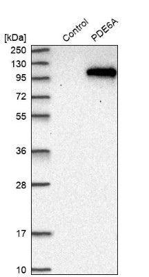
- Western Blot: PDE6A Antibody [NBP1-87312] - Analysis in control (vector only transfected HEK293T lysate) and PDE6A over-expression lysate (Co-expressed with a C-terminal myc-DDK tag (3.1 kDa) in mammalian HEK293T cells).
Supportive validation
- Submitted by
- Novus Biologicals (provider)
- Main image

- Experimental details
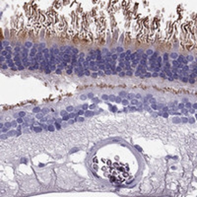
- Immunohistochemistry-Paraffin: PDE6A Antibody [NBP1-87312] - Staining of human retina shows moderate cytoplasmic positivity in rods.
- Submitted by
- Novus Biologicals (provider)
- Main image

- Experimental details
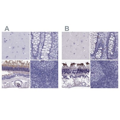
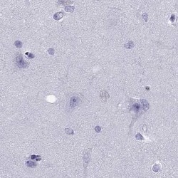
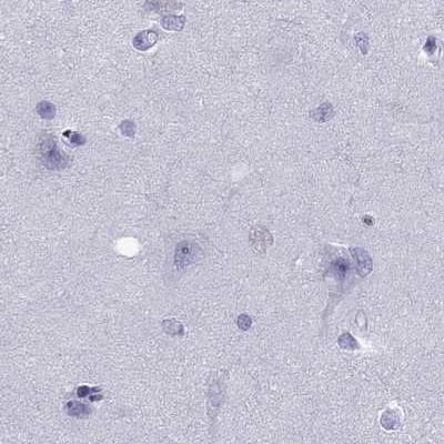
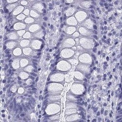
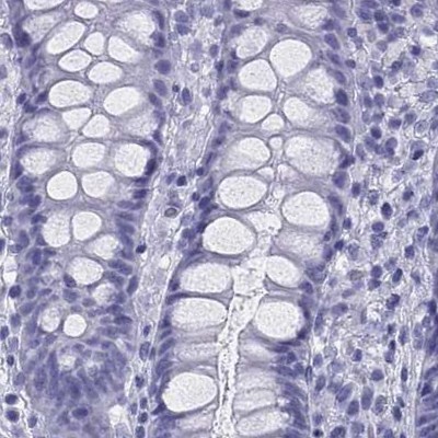
- Immunohistochemistry-Paraffin: PDE6A Antibody [NBP1-87312] - Staining of human cerebral cortex, colon, eye, retina and lymph node using Anti-PDE6A antibody NBP1-87312 (A) shows similar protein distribution across tissues to independent antibody NBP2-68985 (B).
- Submitted by
- Novus Biologicals (provider)
- Main image
- Experimental details
- Immunohistochemistry-Paraffin: PDE6A Antibody [NBP1-87312] - Staining of human cerebral cortex.
- Submitted by
- Novus Biologicals (provider)
- Main image
- Experimental details
- Immunohistochemistry-Paraffin: PDE6A Antibody [NBP1-87312] - Staining of human colon.
- Submitted by
- Novus Biologicals (provider)
- Main image
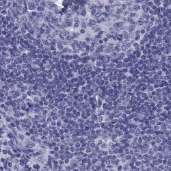
- Experimental details
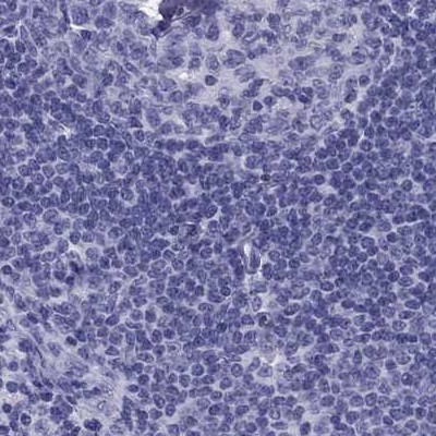
- Immunohistochemistry-Paraffin: PDE6A Antibody [NBP1-87312] - Staining of human lymph node.