 Explore
 Validate
 Learn
Learn Western blot
 Immunocytochemistry
Antibody data
- Antibody Data
- Antigen structure
- References [0]
- Comments [0]
- Validations
- Western blot [1]
- Immunocytochemistry [3]
- Immunohistochemistry [1]
Submit
Validation data
Reference
Comment
Report error
- Product number
- LS-B10995 - Provider product page

- Provider
- LSBio
- Product name
- IHC-plus™ LGALS3 / Galectin 3 Antibody (clone 52C1) LS-B10995
- Antibody type
- Monoclonal
- Description
- Affinity purified
- Reactivity
- Human, Mouse, Rat
- Host
- Mouse
- Isotype
- IgG
- Antibody clone number
- 52C1
- Storage
- Short term: store at 4°C. Long term: aliquot and store at -20°C. Avoid freeze-thaw cycles. Store undiluted.
No comments: Submit comment
Enhanced validation
- Submitted by
- LSBio (provider)
- Enhanced method
- Genetic validation
- Main image
- Experimental details
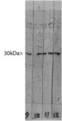
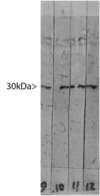
- Blots of crude HeLa cell extract stained with a panel of monoclonal antibodies to Galectin-3. Lane 12 was probed with LGALS3 / Galectin 3 Antibody, revealing a band at the expected molecular weight of 30kDa.
Supportive validation
- Submitted by
- LSBio (provider)
- Enhanced method
- Genetic validation
- Main image

- Experimental details
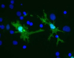
- Rat brain neural cultures stained with LGALS3 / Galectin 3 Antibody (green) and DNA (blue). Staining can be seen in several types of glia and lymphocytic cells, including these cells which have the morphology of microglia. Surrounding cells reveal no Galectin-3 staining.

- Submitted by
- LSBio (provider)
- Main image
- Experimental details
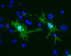
- Rat brain neural cultures stained with LGALS3 / Galectin 3 Antibody (green) and DNA (blue). Staining can be seen in several types of glia and lymphocytic cells, including these cells which have the morphology of microglia. Surrounding cells reveal no Galectin-3 staining.

- Submitted by
- LSBio (provider)
- Main image
- Experimental details
- Rat brain neural cultures stained with LGALS3 / Galectin 3 Antibody (green) and DNA (blue). Staining can be seen in several types of glia and lymphocytic cells, including these cells which have the morphology of microglia. Surrounding cells reveal no Galectin-3 staining.

Supportive validation
- Submitted by
- LSBio (provider)
- Enhanced method
- Genetic validation
- Main image
- Experimental details
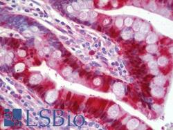
- Human Small Intestine: Formalin-Fixed, Paraffin-Embedded (FFPE)
