Explore
 Validate
 Learn
Learn Western blot
Antibody data
- Antibody Data
- Antigen structure
- References [1]
- Comments [0]
- Validations
- Western blot [6]
- ELISA [1]
- Immunocytochemistry [1]
- Immunohistochemistry [1]
- Other assay [1]
Submit
Validation data
Reference
Comment
Report error
- Product number
- PA5-34819 - Provider product page

- Provider
- Invitrogen Antibodies
- Product name
- Galectin 3 Polyclonal Antibody
- Antibody type
- Polyclonal
- Antigen
- Recombinant protein fragment
- Description
- Recommended positive controls: HeLa, A549, H1299, HCT116, MCF-7, Recombinant LGALS3 protein, rat colon.
- Concentration
- 0.85 mg/mL
Submitted references Endoglin Protein Interactome Profiling Identifies TRIM21 and Galectin-3 as New Binding Partners.
Gallardo-Vara E, Ruiz-Llorente L, Casado-Vela J, Ruiz-Rodríguez MJ, López-Andrés N, Pattnaik AK, Quintanilla M, Bernabeu C
Cells 2019 Sep 13;8(9)
Cells 2019 Sep 13;8(9)
No comments: Submit comment
Supportive validation
- Submitted by
- Invitrogen Antibodies (provider)
- Main image
- Experimental details
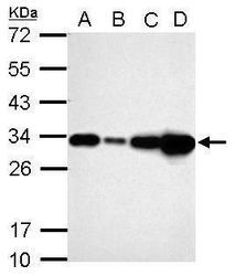
- Western blot analysis of Galectin 3 using 30 µg of A) A549 (B) H1299 (C) HCT116 and D) MCF-7 lysate. Samples were loaded onto a 12% SDS-PAGE gel and probed with a Galectin 3 polyclonal antibody (Product # PA5-34819) at a dilution of 1:10,000.
- Submitted by
- Invitrogen Antibodies (provider)
- Main image
- Experimental details
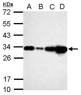
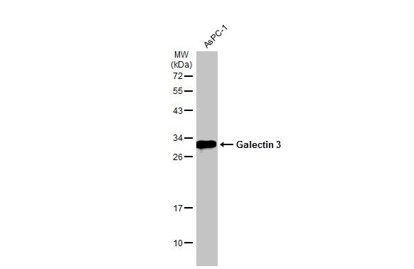
- Western Blot analysis of Galectin 3 was performed by separating 30 µg of various whole cell extracts by 12% SDS-PAGE. Proteins were transferred to a membrane and probed with a Galectin 3 Polyclonal Antibody (Product # PA5-34819) at a dilution of 1:1000 and a HRP-conjugated anti-rabbit IgG secondary antibody.

- Submitted by
- Invitrogen Antibodies (provider)
- Main image
- Experimental details
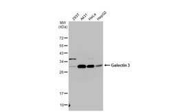
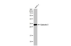
- Western blot analysis of Galectin 3 was performed by separating 30 µg of whole cell extract by 12% SDS-PAGE. Proteins were transferred to a membrane and probed with a Galectin 3 Polyclonal Antibody (Product # PA5-34819) at a dilution of 1:1000. The HRP-conjugated anti-rabbit IgG antibody was used to detect the primary antibody.
- Submitted by
- Invitrogen Antibodies (provider)
- Main image

- Experimental details
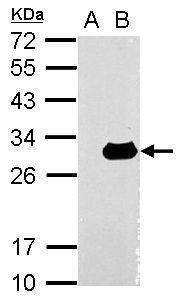
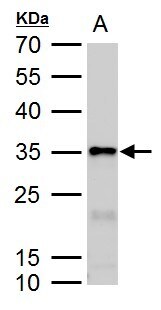
- Western Blot using Galectin 3 Polyclonal Antibody (Product # PA5-34819). Sample (30 µg of whole cell lysate). Lane A: Non-transfected 293T lysates. Lane B: LGALS3 transfected 293T lysates. 12% SDS PAGE. Galectin 3 Polyclonal Antibody (Product # PA5-34819) diluted at 1:10,000.
- Submitted by
- Invitrogen Antibodies (provider)
- Main image
- Experimental details
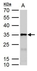
- Galectin3 antibody detects Galectin3 protein by western blot analysis. A. 50 µg rat colon lysate/extract.12 % SDS-PAGE. Galectin3 antibody Galectin 3 Polyclonal Antibody (Product # PA5-34819) dilution: 1:10,000.
- Submitted by
- Invitrogen Antibodies (provider)
- Main image

- Experimental details
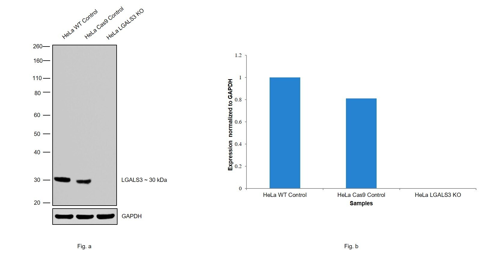
- Knockout of LGALS3 was achieved by CRISPR-Cas9 genome editing using LentiArray™ Lentiviral sgRNA (Product # A32042, AssayID CRISPR855199_LV and CRISPR855204_LV) and LentiArray Cas9 Lentivirus (Product # A32064). Western blot analysis of LGALS3 was performed by loading 30 µg of HeLa wild type (Lane 1), HeLa CAS9 (Lane 2), HeLa LGALS3 KO (Lane 3) membrane extracts. The samples were electrophoresed using Novex® NuPAGE® 4-12% Bis-Tris Protein Gel (Product # NP0321BOX). Resolved proteins were then transferred onto a nitrocellulose membrane (Product # IB23001) by iBlot® 2 Dry Blotting System (Product # IB21001). The blot was probed with Anti-Galectin 3 Polyclonal Antibody (Product # PA5-34819) using 1:1000 dilution and Goat anti-Rabbit IgG (H+L), Superclonal™ Recombinant Secondary Antibody, HRP (Product # A27036, 1:4000 dilution) using the iBright FL 1000 (Product # A32752). Chemiluminescent detection was performed using Novex® ECL Chemiluminescent Substrate Reagent Kit (Product # WP20005). Loss of signal upon CRISPR mediated knockout (KO) using the LentiArray™ CRISPR product line confirms that antibody is specific to LGALS3.
Supportive validation
- Submitted by
- Invitrogen Antibodies (provider)
- Main image
- Experimental details
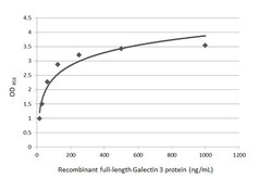
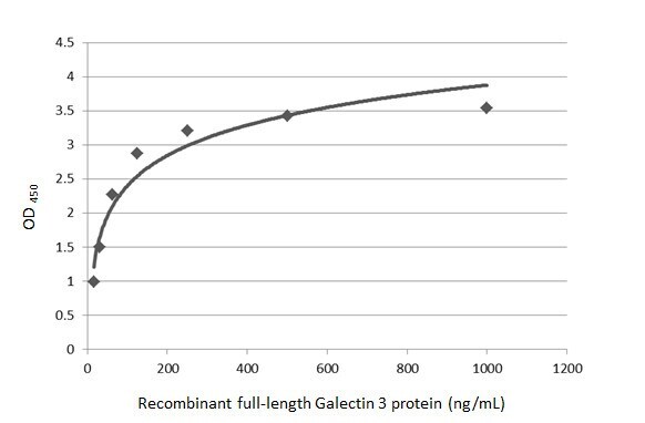
- Sandwich ELISA detection of recombinant full-length Galectin 3 protein using a Galectin 3 Monoclonal Antibody as capture antibody at concentration of 5 µg/mL and Galectin 3 Polyclonal Antibody (Product # PA5-34819) as detection antibody at concentration of 1 µg/mL. Rabbit IgG antibody (HRP) was diluted at 1:10,000 and used to detect the primary antibody.
Supportive validation
- Submitted by
- Invitrogen Antibodies (provider)
- Main image
- Experimental details
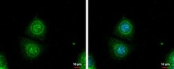
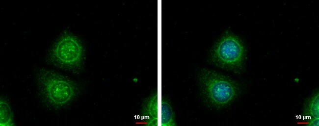
- Galectin3 antibody detects Galectin3 protein at cytoplasm by immunofluorescent analysis. Sample: MCF-7 cells were fixed in ice-cold MeOH for 5 min. Green: Galectin3 protein stained by Galectin3 antibody (Product # PA5-34819) diluted at 1:1,000. Blue: Hoechst 33342 staining. Scale bar = 10 μm.
Supportive validation
- Submitted by
- Invitrogen Antibodies (provider)
- Main image

- Experimental details
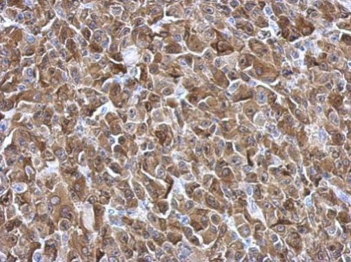
- Immunohistochemical analysis of paraffin-embedded HBL435 xenograft, using Galectin3 (Product # PA5-34819) antibody at 1:500 dilution. Antigen Retrieval: EDTA based buffer, pH 8.0, 15 min.
Supportive validation
- Submitted by
- Invitrogen Antibodies (provider)
- Main image
- Experimental details
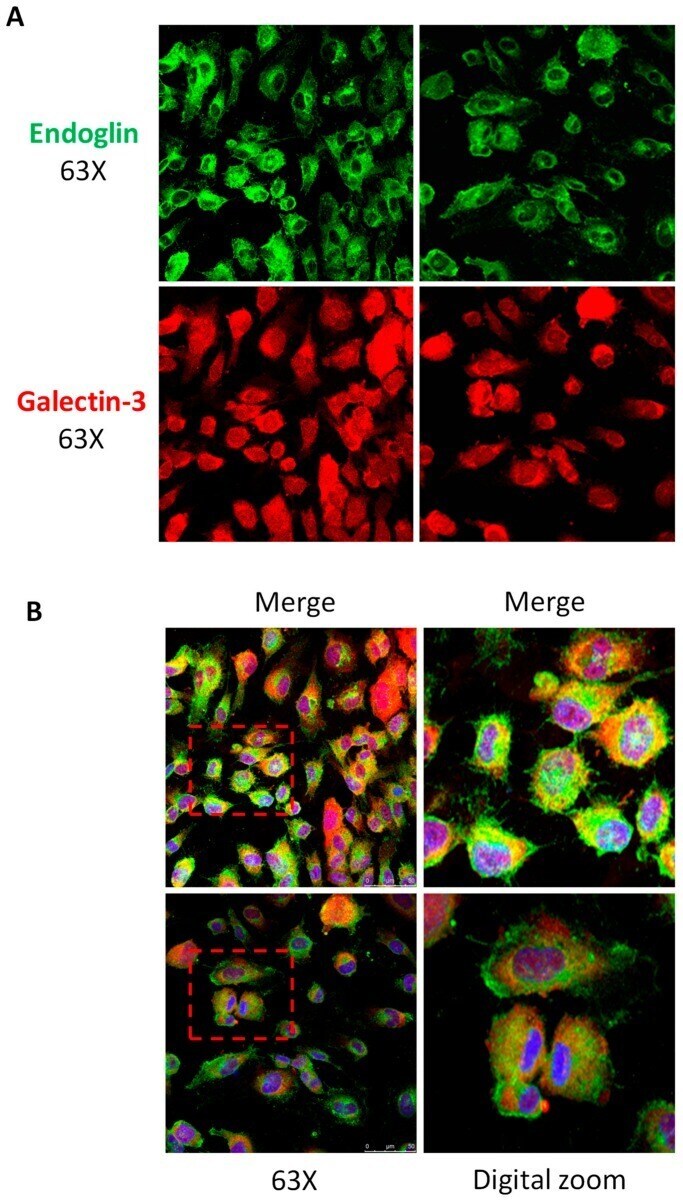
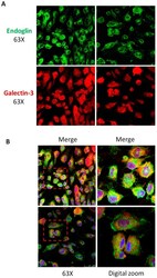
- Figure 2 Galectin-3 and endoglin co-localize in human endothelial cells. Human umbilical vein-derived endothelial cell (HUVEC) monolayers were fixed with paraformaldehyde, permeabilized with Triton X-100, incubated with the mouse mAb P4A4 anti-endoglin, washed, and incubated with a rabbit polyclonal anti-galectin-3 antibody (PA5-34819). Galectin-3 and endoglin were detected by immunofluorescence upon incubation with Alexa 647 goat anti-rabbit IgG (red staining) and Alexa 488 goat anti-mouse IgG (green staining) secondary antibodies, respectively. ( A ) Single staining of galectin-3 (red) and endoglin (green) at the indicated magnifications. ( B ) Merge images plus DAPI (nuclear staining in blue) show co-localization of galectin-3 and endoglin (yellow color). Representative images of five different experiments are shown.