 Explore
 Validate
 Learn
Learn Western blot
 ELISA
ELISAAntibody data
- Antibody Data
- Antigen structure
- References [0]
- Comments [0]
- Validations
- Western blot [2]
- Immunohistochemistry [1]
Submit
Validation data
Reference
Comment
Report error
- Product number
- MAB11541-100 - Provider product page

- Provider
- R&D Systems
- Product name
- Human Galectin-3 Antibody
- Antibody type
- Monoclonal
- Description
- Protein A or G purified from hybridoma culture supernatant. Detects human Galectin-3 in ELISAs. In sandwich immunoassays, no cross-reactivity with recombinant human Galectin-1, 2, 4, 7, 8, or 10 is observed.
- Reactivity
- Human
- Host
- Mouse
- Conjugate
- Unconjugated
- Antigen sequence
P17931.5- Isotype
- IgG
- Antibody clone number
- 194805
- Vial size
- 100 ug
- Concentration
- LYOPH
- Storage
- Use a manual defrost freezer and avoid repeated freeze-thaw cycles. 12 months from date of receipt, -20 to -70 °C as supplied. 1 month, 2 to 8 °C under sterile conditions after reconstitution. 6 months, -20 to -70 °C under sterile conditions after reconstitution.
No comments: Submit comment
Supportive validation
- Submitted by
- R&D Systems (provider)
- Main image
- Experimental details
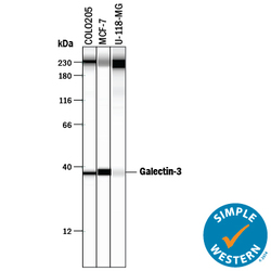
- Detection of Human Galectin-3 by Simple Western<SUP abp="263">TM. Simple Western lane view shows lysates of COLO 205 human colorectal adenocarcinoma cell line, MCF-7 human breast cancer cell line, and U-118-MG human glioblastoma/astrocytoma cell line, loaded at 0.2 mg/mL. A specific band was detected for Galectin-3 at approximately 38 kDa (as indicated) using 10 µg/mL of Mouse Anti-Human Galectin-3 Monoclonal Antibody (Catalog # MAB11541) . This experiment was conducted under reducing conditions and using the 12-230 kDa separation system. Non-specific interaction with the 230 kDa Simple Western standard may be seen with this antibody.

- Submitted by
- R&D Systems (provider)
- Main image
- Experimental details
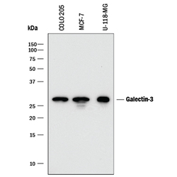
- Detection of Human Galectin-3 by Western Blot. Western blot shows lysates of COLO 205 human colorectal adenocarcinoma cell line, MCF-7 human breast cancer cell line, and U-118-MG human glioblastoma/astrocytoma cell line. PVDF membrane was probed with 0.2 µg/mL of Mouse Anti-Human Galectin-3 Monoclonal Antibody (Catalog # MAB11541) followed by HRP-conjugated Anti-Mouse IgG Secondary Antibody (Catalog # HAF018). A specific band was detected for Galectin-3 at approximately 28 kDa (as indicated). This experiment was conducted under reducing conditions and using Immunoblot Buffer Group 1.

Supportive validation
- Submitted by
- R&D Systems (provider)
- Main image

- Experimental details
- Galectin-3 in Human Prostate Cancer Tissue. Galectin-3 was detected in immersion fixed paraffin-embedded sections of human prostate cancer tissue using Mouse Anti-Human Galectin-3 Monoclonal Antibody (Catalog # MAB11541) at 1.7 µg/mL for 1 hour at room temperature followed by incubation with the Anti-Mouse IgG VisUCyte™ HRP Polymer Antibody (Catalog # VC001). Tissue was stained using DAB (brown) and counterstained with hematoxylin (blue). Specific staining was localized to cytoplasm and plasma membrane in epithelial cells. View our protocol for IHC Staining with VisUCyte HRP Polymer Detection Reagents.
