 Explore
 Validate
 Learn
Learn Western blot
 ELISA
ELISAAntibody data
- Antibody Data
- Antigen structure
- References [3]
- Comments [0]
- Validations
- Western blot [3]
Submit
Validation data
Reference
Comment
Report error
- Product number
- MAB1197 - Provider product page

- Provider
- R&D Systems
- Product name
- Human/Mouse/Rat Galectin-3 Antibody
- Antibody type
- Monoclonal
- Description
- Protein A or G purified from hybridoma culture supernatant. Detects human, mouse, and rat Galectin-3 in Western blots. Detects human and mouse Galectin-3 in direct ELISAs. In direct ELISAs, no cross-reactivity with rhGalectin-2, -4, -8 or recombinant mouse Galectin-1, -4, or -7 is observed.
- Reactivity
- Human, Mouse, Rat
- Host
- Rat
- Conjugate
- Unconjugated
- Antigen sequence
P16110- Isotype
- IgG
- Antibody clone number
- 202213
- Vial size
- 500 ug
- Concentration
- LYOPH
- Storage
- Use a manual defrost freezer and avoid repeated freeze-thaw cycles. 12 months from date of receipt, -20 to -70 °C as supplied. 1 month, 2 to 8 °C under sterile conditions after reconstitution. 6 months, -20 to -70 °C under sterile conditions after reconstitution.
Submitted references Proteomic and functional analysis identifies galectin-1 as a novel regulatory component of the cytotoxic granule machinery.
Galectin-3 negatively regulates the frequency and function of CD4(+) CD25(+) Foxp3(+) regulatory T cells and influences the course of Leishmania major infection.
The receptor of advanced glycation end products plays a central role in advanced oxidation protein products-induced podocyte apoptosis.
Clemente T, Vieira NJ, Cerliani JP, Adrain C, Luthi A, Dominguez MR, Yon M, Barrence FC, Riul TB, Cummings RD, Zorn TM, Amigorena S, Dias-Baruffi M, Rodrigues MM, Martin SJ, Rabinovich GA, Amarante-Mendes GP
Cell death & disease 2017 Dec 7;8(12):e3176
Cell death & disease 2017 Dec 7;8(12):e3176
Galectin-3 negatively regulates the frequency and function of CD4(+) CD25(+) Foxp3(+) regulatory T cells and influences the course of Leishmania major infection.
Fermino ML, Dias FC, Lopes CD, Souza MA, Cruz ÂK, Liu FT, Chammas R, Roque-Barreira MC, Rabinovich GA, Bernardes ES
European journal of immunology 2013 Jul;43(7):1806-17
European journal of immunology 2013 Jul;43(7):1806-17
The receptor of advanced glycation end products plays a central role in advanced oxidation protein products-induced podocyte apoptosis.
Zhou LL, Cao W, Xie C, Tian J, Zhou Z, Zhou Q, Zhu P, Li A, Liu Y, Miyata T, Hou FF, Nie J
Kidney international 2012 Oct;82(7):759-70
Kidney international 2012 Oct;82(7):759-70
No comments: Submit comment
Supportive validation
- Submitted by
- R&D Systems (provider)
- Main image
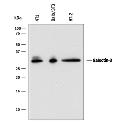
- Experimental details
- Detection of Mouse Galectin-3 by Western Blot. Western blot shows lysates of 4T1 mouse breast cancer cell line, Balb/3T3 mouse embryonic fibroblast cell line and HT-2 mouse T cell line. PVDF membrane was probed with 0.25 µg/mL of Rat Anti-Mouse Galectin-3 Monoclonal Antibody (Catalog # MAB1197) followed by HRP-conjugated Anti-Rat IgG Secondary Antibody (Catalog # HAF005). A specific band was detected for Galectin-3 at approximately 28 kDa (as indicated). This experiment was conducted under reducing conditions and using Immunoblot Buffer Group 1.

- Submitted by
- R&D Systems (provider)
- Main image
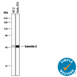
- Experimental details
- Detection of Mouse Galectin-3 by Simple WesternTM. Simple Western lane view shows lysates of HT-2 mouse T cell line and Balb/3T3 mouse embryonic fibroblast cell line, loaded at 0.2 mg/mL. A specific band was detected for Galectin-3 at approximately 43 kDa (as indicated) using 10 µg/mL of Rat Anti-Mouse Galectin-3 Monoclonal Antibody (Catalog # MAB1197) followed by 1:50 dilution of HRP-conjugated Anti-Rat IgG Secondary Antibody (Catalog # HAF005). This experiment was conducted under reducing conditions and using the 12-230 kDa separation system.

- Submitted by
- R&D Systems (provider)
- Main image
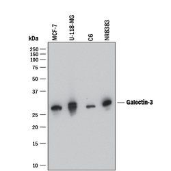
- Experimental details
- Detection of Human and Rat Galectin-3 by Western Blot. Western blot shows lysates of MCF-7 human breast cancer cell line, U-118-MG human glioblastoma/astrocytoma cell line, C6 rat glioma cell line, and NR8383 rat alveolar macrophage cell line. PVDF membrane was probed with 0.5 µg/mL of Rat Anti-Mouse Galectin-3 Monoclonal Antibody (Catalog # MAB1197) followed by HRP-conjugated Anti-Rat IgG Secondary Antibody (Catalog # HAF005). A specific band was detected for Galectin-3 at approximately 28 kDa (as indicated). This experiment was conducted under reducing conditions and using Immunoblot Buffer Group 1.
