Explore
 Validate
 Learn
Learn Flow cytometry
Antibody data
- Antibody Data
- Antigen structure
- References [4]
- Comments [0]
- Validations
- Flow cytometry [1]
- Other assay [4]
Submit
Validation data
Reference
Comment
Report error
- Product number
- 12-5301-82 - Provider product page

- Provider
- Invitrogen Antibodies
- Product name
- Galectin 3 Monoclonal Antibody (eBioM3/38 (M3/38)), PE, eBioscience™
- Antibody type
- Monoclonal
- Antigen
- Other
- Description
- Description: The eBioM3/38 antibody reacts with mouse Galectin-3 (Mac-2). Galectins are a family of animal lectins which appear to exhibit a variety of biological functions. Galectin 3 (aka LGALS3, galactose-specific soluble lectin 3, Mac-2, L-29) is one of the more extensively studied members of this family and is a 30 kDa beta-galactoside-binding protein. Due to a C-terminal carbohydrate binding site, Galectin 3 is capable of binding IgE and mammalian cell surfaces only when homodimerized or homo-oligomerized. Galectin 3 is normally distributed in epithelia of many organs and various inflammatory cells, including macrophages, as well as dendritic cells and Kupffer cells. The expression of this lectin is up-regulated during inflammation, cell proliferation, cell differentiation and through trans-activation by viral proteins.
- Conjugate
- Yellow dye
- Antibody clone number
- eBioM3/38 (M3/38)
- Concentration
- 0.2 mg/mL
Submitted references Endocytic membrane repair by ESCRT-III controls antigen export to the cytosol during antigen cross-presentation.
Cell surface galectin-3 defines a subset of chemoresistant gastrointestinal tumor-initiating cancer cells with heightened stem cell characteristics.
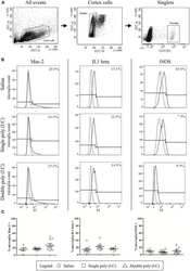
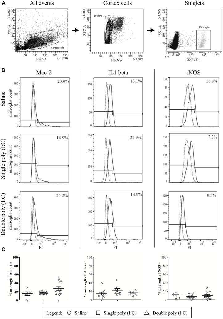
Maternal immune activation evoked by polyinosinic:polycytidylic acid does not evoke microglial cell activation in the embryo.
Galectin-9 controls the therapeutic activity of 4-1BB-targeting antibodies.
Gros M, Segura E, Rookhuizen DC, Baudon B, Heurtebise-Chrétien S, Burgdorf N, Maurin M, Kapp EA, Simpson RJ, Kozik P, Villadangos JA, Bertrand MJM, Burbage M, Amigorena S
Cell reports 2022 Aug 16;40(7):111205
Cell reports 2022 Aug 16;40(7):111205
Cell surface galectin-3 defines a subset of chemoresistant gastrointestinal tumor-initiating cancer cells with heightened stem cell characteristics.
Ilmer M, Mazurek N, Byrd JC, Ramirez K, Hafley M, Alt E, Vykoukal J, Bresalier RS
Cell death & disease 2016 Aug 11;7(8):e2337
Cell death & disease 2016 Aug 11;7(8):e2337
Maternal immune activation evoked by polyinosinic:polycytidylic acid does not evoke microglial cell activation in the embryo.
Smolders S, Smolders SM, Swinnen N, Gärtner A, Rigo JM, Legendre P, Brône B
Frontiers in cellular neuroscience 2015;9:301
Frontiers in cellular neuroscience 2015;9:301
Galectin-9 controls the therapeutic activity of 4-1BB-targeting antibodies.
Madireddi S, Eun SY, Lee SW, Nemčovičová I, Mehta AK, Zajonc DM, Nishi N, Niki T, Hirashima M, Croft M
The Journal of experimental medicine 2014 Jun 30;211(7):1433-48
The Journal of experimental medicine 2014 Jun 30;211(7):1433-48
No comments: Submit comment
Supportive validation
- Submitted by
- Invitrogen Antibodies (provider)
- Main image

- Experimental details
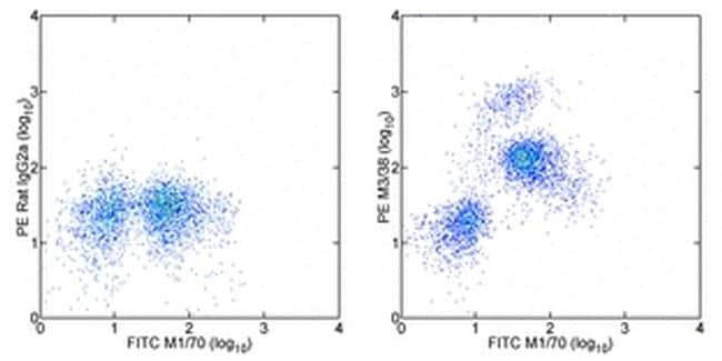
- Staining of C57BL/6 bone marrow cells with Anti-Mouse CD11b FITC (Product # 11-0112-41) and Rat IgG2a K Isotype Control PE (Product # 12-4321-80) (left) or 0.03 µg of Anti-Human/Mouse Galectin-3 PE (right). Total viable cells were used for analysis.
- Conjugate
- Yellow dye
Supportive validation
- Submitted by
- Invitrogen Antibodies (provider)
- Main image
- Experimental details
- NULL
- Conjugate
- Yellow dye
- Submitted by
- Invitrogen Antibodies (provider)
- Main image

- Experimental details
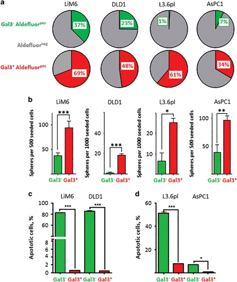
- Figure 2 Gal3 positive CSC subset displays increased stemness characteristics. ( a ) ALDH activity was evaluated by flow cytometric analysis in Gal3 positive CSC (in red) and Gal3 negative CSC (in green). ALDH negative fractions are illustrated in gray. ( b ) Sphere formation ability (SFA) was determined. Gal3 positive CSC (red) accounted for significantly more spheres than Gal3 negative CSC (green). ( c ) TRAIL-resistance in Gal3 positive /Gal3 negative colon cancer CSC subsets was determined by APO-BRDU assay. Apoptotic cells are displayed as percentage of total cell count. ( d ) TRAIL-resistance in Gal3 positive /Gal3 negative CSC pancreatic cancer subsets. All experiments were performed in triplicate (see text for details). P
- Conjugate
- Yellow dye
- Submitted by
- Invitrogen Antibodies (provider)
- Main image
- Experimental details
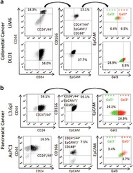
- Figure 1 Surface Gal3 defines a subtype of epithelial CSC. ( a ) Colorectal cancer cells LiM6 and DLD1 were investigated by flow cytometry for cell surface markers CD24/CD44 (left). CD24+/CD44+-cells (in black) were then investigated for CD166/EpCAM-expression (middle) and CD24+/CD44+/CD166+/EpCAM+-positive cells (in black middle) for Gal3-expression (right panel), where ++++ Gal3 - in green refers to cells positive for CD24, CD44, CD166, and EpCAM, but negative for Gal3, and ++++ Gal3 + in red refers to cells positive for CD24, CD44, CD166, EpCAM, and Gal3. ( b ) Pancreatic cancer cell lines AsPC1 and L3.6pl were subjected to the same analysis as in ( a )
- Conjugate
- Yellow dye

- Submitted by
- Invitrogen Antibodies (provider)
- Main image
- Experimental details
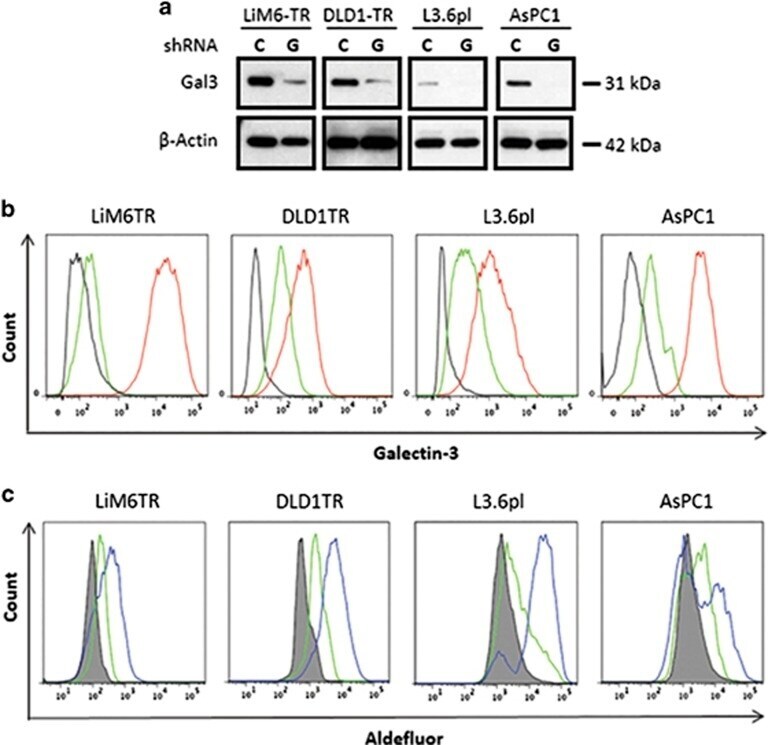
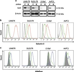
- Figure 4 Silencing of Gal3 shifts CSC subset to a Gal3 negative CSC subset. ( a ) Western blot analysis for comparison of total Gal3 in LiM6TR or DLD1TR colon cancer cells and L3.6pl or AsPC1 pancreatic cancer cells after infection with lentiviral particles for control-shRNA ( c ) or Gal3-shRNA (G). Each pair of control and Gal3 knockdown cells were run side by side on the same gel. Comparison of Gal3 expression between cell lines has been previously published 10 ( b ) Flow cytometric analysis of colon cancer and pancreatic cancer cells after infection with lentiviral particles for control-shRNA (red trace) or Gal3-shRNA (green trace). Black trace is background staining. ( c ) ALDH activity was analyzed by flow cytometry. ALDEFLUOR activity in colon cancer and pancreatic cancer cells after infection with lentiviral particles for control-shRNA (blue trace) or Gal3-shRNA (green trace). Gray profile is background staining
- Conjugate
- Yellow dye