Explore
 Validate
 Learn
Learn Flow cytometry
Antibody data
- Antibody Data
- Antigen structure
- References [5]
- Comments [0]
- Validations
- Flow cytometry [1]
Submit
Validation data
Reference
Comment
Report error
- Product number
- 50-5301-80 - Provider product page

- Provider
- Invitrogen Antibodies
- Product name
- Anti-Galectin 3 Monoclonal Antibody (eBioM3/38 (M3/38)), eFluor 660, eBioscience™
- Antibody type
- Monoclonal
- Antigen
- Other
- Description
- Description: The eBioM3/38 (M3/38) antibody reacts with mouse Galectin-3 (Mac-2). Galectins are a family of animal lectins which appear to exhibit a variety of biological functions. Galectin 3 (aka LGALS3, galactose-specific soluble lectin 3, Mac-2, L-29) is one of the more extensively studied members of this family and is a 30 kDa beta-galactoside-binding protein. Due to a C-terminal carbohydrate binding site, Galectin 3 is capable of binding IgE and mammalian cell surfaces only when homodimerized or homo-oligomerized. Galectin 3 is normally distributed in epithelia of many organs and various inflammatory cells, including macrophages, as well as dendritic cells and Kupffer cells. The expression of this lectin is up-regulated during inflammation, cell proliferation, cell differentiation and through trans-activation by viral proteins. This monoclonal antibody eBioM3/38 crossreacts to human Galectin-3 (LGALS3). Applications Reported: This eBioM3/38 (M3/38) antibody has been reported for use in flow cytometric analysis. Applications Tested: This eBioM3/38 (M3/38) antibody has been tested by flow cytometric analysis of mouse bone marrow cells. This can be used at less than or equal to 0.125 µg per test. A test is defined as the amount (µg) of antibody that will stain a cell sample in a final volume of 100 µL. Cell number should be determined empirically but can range from 10^5 to 10^8 cells/test. It is recommended that the antibody be carefully titrated for optimal performance in the assay of interest. eFluor® 660 is a replacement for Alexa Fluor® 647. eFluor® 660 emits at 659 nm and is excited with the red laser (633 nm). Please make sure that your instrument is capable of detecting this fluorochome. Excitation: 633-647 nm; Emission: 668 nm; Laser: Red Laser. Filtration: 0.2 µm post-manufacturing filtered.
- Reactivity
- Human, Mouse
- Host
- Rat
- Isotype
- IgG
- Antibody clone number
- eBioM3/38 (M3/38)
- Vial size
- 25 µg
- Concentration
- 0.2 mg/mL
- Storage
- 4° C, store in dark, DO NOT FREEZE!
Submitted references Mixed-lineage kinase 3 pharmacological inhibition attenuates murine nonalcoholic steatohepatitis.
Galectin-3 expression in hippocampal CA2 following transient forebrain ischemia and its inhibition by hypothermia or antiapoptotic agents.
Cell surface galectin-3 defines a subset of chemoresistant gastrointestinal tumor-initiating cancer cells with heightened stem cell characteristics.
Maternal immune activation evoked by polyinosinic:polycytidylic acid does not evoke microglial cell activation in the embryo.
Imaging white adipose tissue with confocal microscopy.
Tomita K, Kohli R, MacLaurin BL, Hirsova P, Guo Q, Sanchez LHG, Gelbard HA, Blaxall BC, Ibrahim SH
JCI insight 2017 Aug 3;2(15)
JCI insight 2017 Aug 3;2(15)
Galectin-3 expression in hippocampal CA2 following transient forebrain ischemia and its inhibition by hypothermia or antiapoptotic agents.
Hisamatsu K, Niwa M, Kobayashi K, Miyazaki T, Hirata A, Hatano Y, Tomita H, Hara A
Neuroreport 2016 Mar 23;27(5):311-7
Neuroreport 2016 Mar 23;27(5):311-7
Cell surface galectin-3 defines a subset of chemoresistant gastrointestinal tumor-initiating cancer cells with heightened stem cell characteristics.
Ilmer M, Mazurek N, Byrd JC, Ramirez K, Hafley M, Alt E, Vykoukal J, Bresalier RS
Cell death & disease 2016 Aug 11;7(8):e2337
Cell death & disease 2016 Aug 11;7(8):e2337
Maternal immune activation evoked by polyinosinic:polycytidylic acid does not evoke microglial cell activation in the embryo.
Smolders S, Smolders SM, Swinnen N, Gärtner A, Rigo JM, Legendre P, Brône B
Frontiers in cellular neuroscience 2015;9:301
Frontiers in cellular neuroscience 2015;9:301
Imaging white adipose tissue with confocal microscopy.
Martinez-Santibañez G, Cho KW, Lumeng CN
Methods in enzymology 2014;537:17-30
Methods in enzymology 2014;537:17-30
No comments: Submit comment
Supportive validation
- Submitted by
- Invitrogen Antibodies (provider)
- Main image
- Experimental details
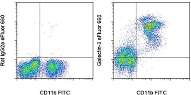
- Staining of C57Bl/6 bone marrow cells with Anti-Mouse CD11b FITC (Product # 11-0112-41) and 0.06 µg of Rat IgG2a K Isotype Control eFluor® 660 (Product # 50-4321-82) (left) or 0.06 µg of Anti-Human/Mouse Galectin-3 eFluor® 660 (right). Cells in the large scatter population were used for analysis.