Explore
 Validate
 Learn
Learn Western blot
Antibody data
- Antibody Data
- Antigen structure
- References [0]
- Comments [0]
- Validations
- Western blot [1]
- Immunocytochemistry [2]
- Immunohistochemistry [2]
- Flow cytometry [1]
Submit
Validation data
Reference
Comment
Report error
- Product number
- NBP2-67160 - Provider product page

- Provider
- Novus Biologicals
- Product name
- Rabbit Monoclonal APLP-1 Antibody
- Antibody type
- Monoclonal
- Description
- Protein A purified.
- Reactivity
- Human, Mouse, Rat
- Host
- Rabbit
- Isotype
- IgG
- Vial size
- 100 ul
- Storage
- Store at 4C short term. Aliquot and store at -20C long term. Avoid freeze-thaw cycles.
No comments: Submit comment
Supportive validation
- Submitted by
- Novus Biologicals (provider)
- Main image
- Experimental details
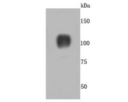
- Western Blot: APLP-1 Antibody (JM11-42) [NBP2-67160] - Analysis of CD26 on mouse heart tissue lysates using anti-CD26 antibody at 1/500 dilution.

Supportive validation
- Submitted by
- Novus Biologicals (provider)
- Main image
- Experimental details
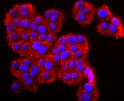
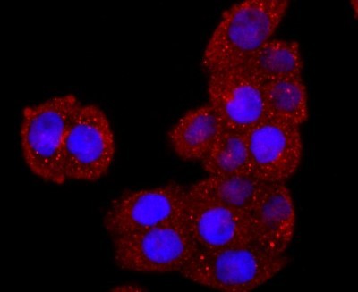
- Immunocytochemistry/Immunofluorescence: APLP-1 Antibody (JM11-42) [NBP2-67160] - Staining CD26 in PC-12 cells (red). The nuclear counter stain is DAPI (blue). Cells were fixed in paraformaldehyde, permeabilised with 0.25% Triton X100/PBS.
- Submitted by
- Novus Biologicals (provider)
- Main image

- Experimental details
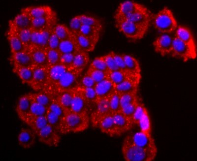
- Immunocytochemistry/Immunofluorescence: APLP-1 Antibody (JM11-42) [NBP2-67160] - Staining CD26 in Hela cells (red). The nuclear counter stain is DAPI (blue). Cells were fixed in paraformaldehyde, permeabilised with 0.25% Triton X100/PBS.
Supportive validation
- Submitted by
- Novus Biologicals (provider)
- Main image
- Experimental details
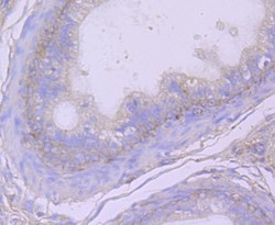
- Immunohistochemistry-Paraffin: APLP-1 Antibody (JM11-42) [NBP2-67160] - Analysis of paraffin-embedded rat epididymis tissue using anti-CD26 antibody. Counter stained with hematoxylin.

- Submitted by
- Novus Biologicals (provider)
- Main image
- Experimental details
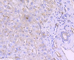
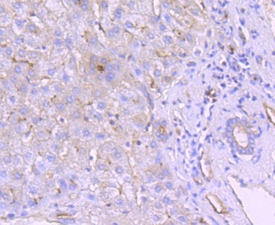
- Immunohistochemistry-Paraffin: APLP-1 Antibody (JM11-42) [NBP2-67160] - Analysis of paraffin-embedded human liver tissue using anti-CD26 antibody. Counter stained with hematoxylin.
Supportive validation
- Submitted by
- Novus Biologicals (provider)
- Main image

- Experimental details
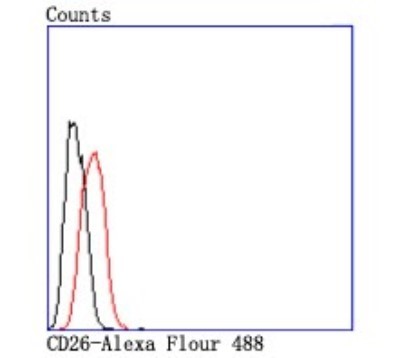
- Flow Cytometry: APLP-1 Antibody (JM11-42) [NBP2-67160] - Analysis of Hela cells with CD26 antibody at 1/50 dilution (red) compared with an unlabelled control (cells without incubation with primary antibody; black). Alexa Fluor 488-conjugated goat anti rabbit IgG was used as the secondary antibody.