 Explore
 Validate
 Learn
Learn Western blot
Antibody data
- Antibody Data
- Antigen structure
- References [8]
- Comments [0]
- Validations
- Western blot [3]
- Immunocytochemistry [1]
- Flow cytometry [1]
Submit
Validation data
Reference
Comment
Report error
- Product number
- MAB6158 - Provider product page

- Provider
- R&D Systems
- Product name
- Human/Mouse UCP1 Antibody
- Antibody type
- Monoclonal
- Description
- Protein A or G purified from hybridoma culture supernatant. Detects human and mouse UCP1 in Western blots.
- Reactivity
- Human, Mouse
- Host
- Mouse
- Conjugate
- Unconjugated
- Antigen sequence
P25874- Isotype
- IgG
- Antibody clone number
- 536435
- Vial size
- 100 ug
- Concentration
- LYOPH
- Storage
- Use a manual defrost freezer and avoid repeated freeze-thaw cycles. 12 months from date of receipt, -20 to -70 °C as supplied. 1 month, 2 to 8 °C under sterile conditions after reconstitution. 6 months, -20 to -70 °C under sterile conditions after reconstitution.
Submitted references Differentiating SGBS adipocytes respond to PPARγ stimulation, irisin and BMP7 by functional browning and beige characteristics.
Mature Human White Adipocytes Cultured under Membranes Maintain Identity, Function, and Can Transdifferentiate into Brown-like Adipocytes.
Flow Cytometry of Mouse and Human Adipocytes for the Analysis of Browning and Cellular Heterogeneity.
A Renewable Source of Human Beige Adipocytes for Development of Therapies to Treat Metabolic Syndrome.
IRX3 Promotes the Browning of White Adipocytes and Its Rare Variants are Associated with Human Obesity Risk.
Both brown adipose tissue and skeletal muscle thermogenesis processes are activated during mild to severe cold adaptation in mice.
Direct conversion of human fibroblasts to brown adipocytes by small chemical compounds.
White-to-brown metabolic conversion of human adipocytes by JAK inhibition.
Klusóczki Á, Veréb Z, Vámos A, Fischer-Posovszky P, Wabitsch M, Bacso Z, Fésüs L, Kristóf E
Scientific reports 2019 Apr 9;9(1):5823
Scientific reports 2019 Apr 9;9(1):5823
Mature Human White Adipocytes Cultured under Membranes Maintain Identity, Function, and Can Transdifferentiate into Brown-like Adipocytes.
Harms MJ, Li Q, Lee S, Zhang C, Kull B, Hallen S, Thorell A, Alexandersson I, Hagberg CE, Peng XR, Mardinoglu A, Spalding KL, Boucher J
Cell reports 2019 Apr 2;27(1):213-225.e5
Cell reports 2019 Apr 2;27(1):213-225.e5
Flow Cytometry of Mouse and Human Adipocytes for the Analysis of Browning and Cellular Heterogeneity.
Hagberg CE, Li Q, Kutschke M, Bhowmick D, Kiss E, Shabalina IG, Harms MJ, Shilkova O, Kozina V, Nedergaard J, Boucher J, Thorell A, Spalding KL
Cell reports 2018 Sep 4;24(10):2746-2756.e5
Cell reports 2018 Sep 4;24(10):2746-2756.e5
A Renewable Source of Human Beige Adipocytes for Development of Therapies to Treat Metabolic Syndrome.
Su S, Guntur AR, Nguyen DC, Fakory SS, Doucette CC, Leech C, Lotana H, Kelley M, Kohli J, Martino J, Sims-Lucas S, Liaw L, Vary C, Rosen CJ, Brown AC
Cell reports 2018 Dec 11;25(11):3215-3228.e9
Cell reports 2018 Dec 11;25(11):3215-3228.e9
IRX3 Promotes the Browning of White Adipocytes and Its Rare Variants are Associated with Human Obesity Risk.
Zou Y, Lu P, Shi J, Liu W, Yang M, Zhao S, Chen N, Chen M, Sun Y, Gao A, Chen Q, Zhang Z, Ma Q, Ning T, Ying X, Jin J, Deng X, Shen B, Zhang Y, Yuan B, Kauderer S, Liu S, Hong J, Liu R, Ning G, Wang W, Gu W, Wang J
EBioMedicine 2017 Oct;24:64-75
EBioMedicine 2017 Oct;24:64-75
Both brown adipose tissue and skeletal muscle thermogenesis processes are activated during mild to severe cold adaptation in mice.
Bal NC, Singh S, Reis FCG, Maurya SK, Pani S, Rowland LA, Periasamy M
The Journal of biological chemistry 2017 Oct 6;292(40):16616-16625
The Journal of biological chemistry 2017 Oct 6;292(40):16616-16625
Direct conversion of human fibroblasts to brown adipocytes by small chemical compounds.
Takeda Y, Harada Y, Yoshikawa T, Dai P
Scientific reports 2017 Jun 27;7(1):4304
Scientific reports 2017 Jun 27;7(1):4304
White-to-brown metabolic conversion of human adipocytes by JAK inhibition.
Moisan A, Lee YK, Zhang JD, Hudak CS, Meyer CA, Prummer M, Zoffmann S, Truong HH, Ebeling M, Kiialainen A, Gérard R, Xia F, Schinzel RT, Amrein KE, Cowan CA
Nature cell biology 2015 Jan;17(1):57-67
Nature cell biology 2015 Jan;17(1):57-67
No comments: Submit comment
Supportive validation
- Submitted by
- R&D Systems (provider)
- Main image
- Experimental details
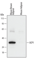
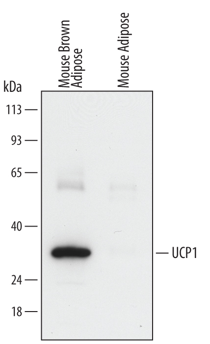
- Detection of Mouse UCP1 by Western Blot. Western blot shows lysates of mouse brown adipose tissue and mouse adipose tissue. PVDF Membrane was probed with 0.5 µg/mL of Mouse Anti-Human/Mouse UCP1 Monoclonal Antibody (Catalog # MAB6158) followed by HRP-conjugated Anti-Mouse IgG Secondary Antibody (Catalog # HAF007). A specific band was detected for UCP1 at approximately 33 kDa (as indicated). This experiment was conducted under reducing conditions and using Immunoblot Buffer Group 2.
- Submitted by
- R&D Systems (provider)
- Main image
- Experimental details
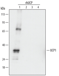
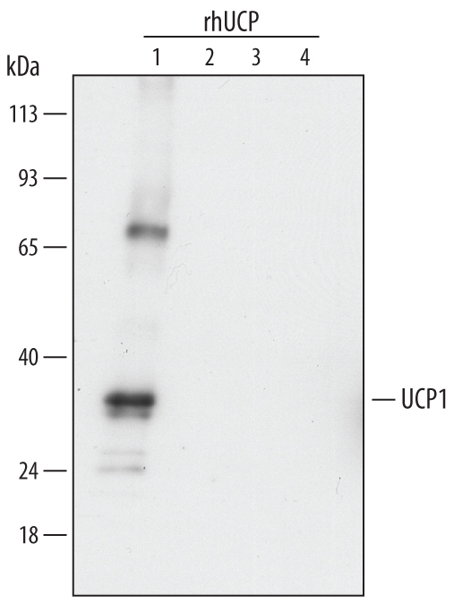
- Detection of Human UCP1 by Western Blot. Western blot shows recombinant human UCP1, recombinant human UCP2, recombinant human UCP3, and recombinant human UCP4 (5 ng/lane). PVDF Membrane was probed with 0.5 µg/mL of Mouse Anti-Human/Mouse UCP1 Monoclonal Antibody (Catalog # MAB6158) followed by HRP-conjugated Anti-Mouse IgG Secondary Antibody (Catalog # HAF007). This experiment was conducted under reducing conditions and using Immunoblot Buffer Group 2.
- Submitted by
- R&D Systems (provider)
- Main image

- Experimental details
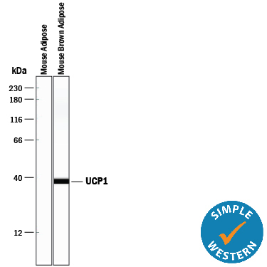
- Detection of Mouse UCP1 by Simple WesternTM. Simple Western lane view shows lysates of mouse adipose tissue and mouse brown adipose tissue, loaded at 0.5 mg/mL. A specific band was detected for UCP1 at approximately 37 kDa (as indicated) using 2.5 µg/mL of Mouse Anti-Human/Mouse UCP1 Monoclonal Antibody (Catalog # MAB6158). This experiment was conducted under reducing conditions and using the 12-230 kDa separation system.
Supportive validation
- Submitted by
- R&D Systems (provider)
- Main image
- Experimental details
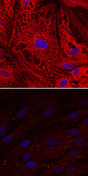
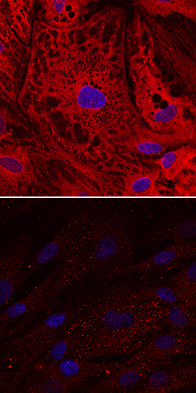
- UCP1 in Human Mesenchymal Stem Cells. UCP1 was detected in immersion fixed human mesenchymal stem cells undifferentiated (lower panel) or differentiated into adipocytes (upper panel) using Mouse Anti-Human/Mouse UCP1 Monoclonal Antibody (Catalog # MAB6158) at 10 µg/mL for 3 hours at room temperature. Cells were stained using the NorthernLights™ 557-conjugated Anti-Mouse IgG Secondary Antibody (red; Catalog # NL007) and counterstained with DAPI (blue). Specific staining was localized to cytoplasm. View our protocol for Fluorescent ICC Staining of Stem Cells on Coverslips.
Supportive validation
- Submitted by
- R&D Systems (provider)
- Main image
- Experimental details
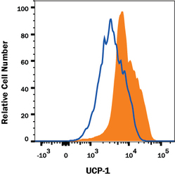
- Detection of UCP1 in 3T3-L1 Mouse Cell Line by Flow Cytometry. 3T3-L1 mouse embryonic fibroblast adipose-like cell line was stained with Mouse Anti-Human/Mouse UCP1 Monoclonal Antibody (Catalog # MAB6158, filled histogram) or isotype control antibody (Catalog # MAB0041, open histogram) followed by anti-Mouse IgG PE-conjugated Secondary Antibody (Catalog # F0102B). To facilitate intracellular staining, cells were fixed with Flow Cytometry Fixation Buffer (Catalog # FC004) and permeabilized with Flow Cytometry Permeabilization/Wash Buffer I (Catalog # FC005). View our protocol for Staining Intracellular Molecules.
