 Explore
 Validate
 Learn
Learn Western blot
Antibody data
- Antibody Data
- Antigen structure
- References [0]
- Comments [0]
- Validations
- Western blot [3]
- Immunocytochemistry [1]
- Immunohistochemistry [1]
Submit
Validation data
Reference
Comment
Report error
- Product number
- PA5-52890 - Provider product page

- Provider
- Invitrogen Antibodies
- Product name
- Anti-Monoacylglycerol Lipase Polyclonal Antibody
- Antibody type
- Polyclonal
- Antigen
- Recombinant full-length protein
- Description
- Immunogen sequence: LVLANPESAT TFKVLAAKVL NLVLPNLSLG PIDSSVLSRN KTEVDIYNSD PLICRAGLKV CFGIQLLNAV SRVERALPKL TVPFLLLQGS ADRLCDSKGA YLLMELAKSQ DKTLKIYEGA YHVLHKELPE VTNSVFHE Highest antigen sequence identity to the following orthologs: Mouse - 86%, Rat - 86%.
- Reactivity
- Human, Mouse, Rat
- Host
- Rabbit
- Isotype
- IgG
- Vial size
- 100 µL
- Concentration
- 0.1 mg/mL
- Storage
- Store at 4°C short term. For long term storage, store at -20°C, avoiding freeze/thaw cycles.
No comments: Submit comment
Supportive validation
- Submitted by
- Invitrogen Antibodies (provider)
- Main image

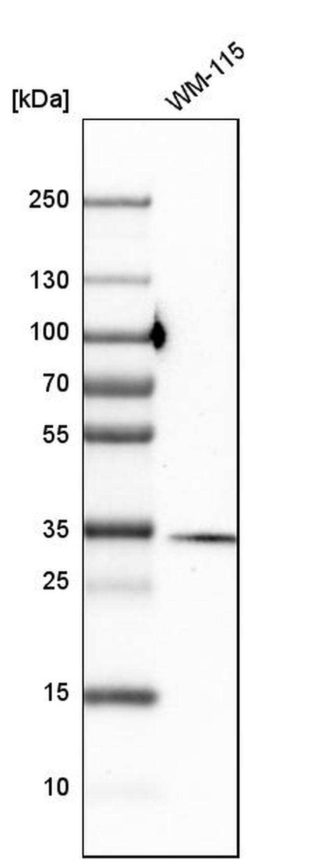
- Experimental details
- Western blot analysis of Monoacylglycerol Lipase in human cell line WM-115 using a Monoacylglycerol Lipase Polyclonal Antibody (Product # PA5-52890).
- Submitted by
- Invitrogen Antibodies (provider)
- Main image

- Experimental details
- Western blot analysis of Monoacylglycerol Lipase in control (vector only transfected HEK293T lysate) and MGLL over-expression lysate (Co-expressed with a C-terminal myc-DDK tag (~3.1 kDa) in mammalian HEK293T cells). Samples were probed using a Monoacylglycerol Lipase Polyclonal Antibody (Product # PA5-52890).

- Submitted by
- Invitrogen Antibodies (provider)
- Main image

- Experimental details
- Western blot was performed using Anti-Monoacylglycerol Lipase Polyclonal Antibody (Product # PA5-52890) and a 35KDa band corresponding to Monoacylglycerol Lipase was observed along with uncharacterized band (*) in tissues tested. Tissue extracts (30 µg lysate) of Mouse Adipose (Lane 1), Mouse Lung (Lane 2), Rat Lung (Lane 3), Mouse liver (Lane 4) and Rat liver (Lane 5) were electrophoresed using NuPAGE™ 4-12% Bis-Tris Protein Gel (Product # NP0322BOX). Resolved proteins were then transferred onto a nitrocellulose membrane (Product # IB23001) by iBlot® 2 Dry Blotting System (Product # IB21001). The blot was probed with the primary antibody (0.4 µg/mL) and detected by chemiluminescence with Goat anti-Rabbit IgG (H+L) Superclonal™ Recombinant Secondary Antibody, HRP (Product # A27036, 1:4000 dilution) using the iBright FL 1000 (Product # A32752). Chemiluminescent detection was performed using Novex® ECL Chemiluminescent Substrate Reagent Kit (Product # WP20005) (http://www.jbc.org/content/272/43/27218.full.pdf).

Supportive validation
- Submitted by
- Invitrogen Antibodies (provider)
- Main image

- Experimental details
- Immunofluorescent staining of Monoglyceride Lipase in human cell line U-251 MG shows positivity in nucleus but excluded from the nucleoli. Samples were probed using a Monoglyceride Lipase Polyclonal Antibody (Product # PA5-52890).

Supportive validation
- Submitted by
- Invitrogen Antibodies (provider)
- Main image

- Experimental details
- Immunohistochemical staining of Monoglyceride Lipase in human colon tissue shows strong cytoplasmic positivity in glandular cells. Samples were probed using a Monoglyceride Lipase Polyclonal Antibody (Product # PA5-52890).
