Explore
 Validate
 Learn
Learn Western blot
 ELISA
ELISA Immunocytochemistry
Antibody data
- Antibody Data
- Antigen structure
- References [2]
- Comments [0]
- Validations
- Immunocytochemistry [3]
- Immunohistochemistry [1]
- Flow cytometry [2]
- Other assay [2]
Submit
Validation data
Reference
Comment
Report error
- Product number
- MA5-15677 - Provider product page

- Provider
- Invitrogen Antibodies
- Product name
- SIRT1 Monoclonal Antibody (1F3)
- Antibody type
- Monoclonal
- Antigen
- Purifed from natural sources
- Description
- MA5-15677 targets SIRT1 in indirect ELISA, FACS, IF, IHC, and WB applications and shows reactivity with Human and Non-human primate samples. The MA5-15677 immunogen is purified recombinant fragment of human SIRT1 expressed in E. Coli. MA5-15677 detects SIRT1 which has a predicted molecular weight of approximately 120kDa.
- Reactivity
- Human
- Host
- Mouse
- Isotype
- IgG
- Antibody clone number
- 1F3
- Vial size
- 100 μL
- Concentration
- Conc. Not Determined
- Storage
- Store at 4°C short term. For long term storage, store at -20°C, avoiding freeze/thaw cycles.
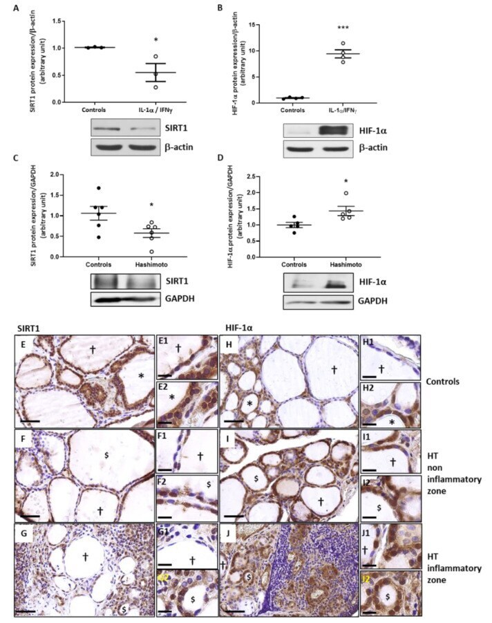
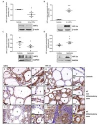
Submitted references Oxidative Stress-Induced Sirtuin1 Downregulation Correlates to HIF-1α, GLUT-1, and VEGF-A Upregulation in Th1 Autoimmune Hashimoto's Thyroiditis.
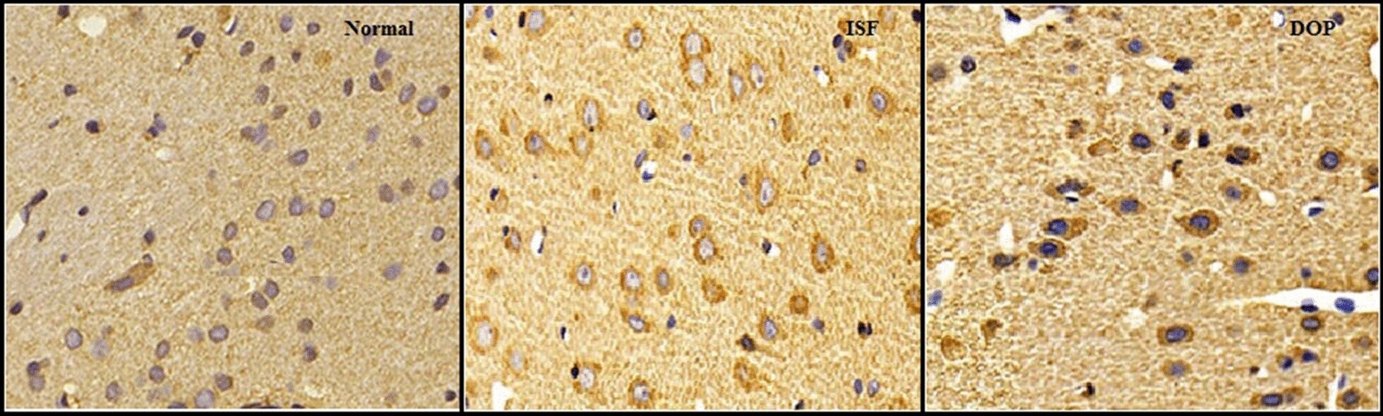
Desoxyrhapontigenin attenuates neuronal apoptosis in an isoflurane-induced neuronal injury model by modulating the TLR-4/cyclin B1/Sirt-1 pathway.
Hepp M, Werion A, De Greef A, de Ville de Goyet C, de Bournonville M, Behets C, Lengelé B, Daumerie C, Mourad M, Ludgate M, Many MC, Joris V, Craps J
International journal of molecular sciences 2021 Apr 7;22(8)
International journal of molecular sciences 2021 Apr 7;22(8)
Desoxyrhapontigenin attenuates neuronal apoptosis in an isoflurane-induced neuronal injury model by modulating the TLR-4/cyclin B1/Sirt-1 pathway.
Liang F, Fu X, Li Y, Han F
AMB Express 2020 Sep 30;10(1):175
AMB Express 2020 Sep 30;10(1):175
No comments: Submit comment
Supportive validation
- Submitted by
- Invitrogen Antibodies (provider)
- Main image
- Experimental details
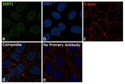
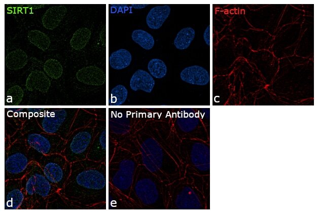
- Immunofluorescence analysis of SIRT1 was performed using 70% confluent log phase NTERA-2 cells. The cells were fixed with 4% paraformaldehyde for 10 minutes, permeabilized with 0.1% Triton™ X-100 for 15 minutes, and blocked with 1% BSA for 1 hour at room temperature. The cells were labeled with SIRT1 Monoclonal Antibody (1F3) (Product # MA5-15677) at 1:250 dilution in 0.1% BSA, incubated at 4 degree Celsius overnight and then labeled with Goat anti-Mouse IgG (H+L) Superclonal™ Secondary Antibody, Alexa Fluor® 488 conjugate (Product # A28175) for 45 minutes at room temperature (Panel a: green). Nuclei (Panel b: blue) were stained with ProLong™ Diamond Antifade Mountant with DAPI (Product # P36962). F-actin (Panel c: red) was stained with Rhodamine Phalloidin (Product # R415, 1:300). Panel d represents the merged image showing nuclear localization of SIRT1. Panel e represents control cells with no primary antibody to assess background. The images were captured at 60X magnification.
- Submitted by
- Invitrogen Antibodies (provider)
- Main image

- Experimental details
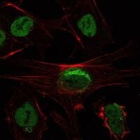
- Immunofluorescence analysis of NTERA-2 cells using SIRT1 monoclonal antibody (Product # MA5-15677) (Green). Red: actin filaments have been labeled with phalloidin.
- Submitted by
- Invitrogen Antibodies (provider)
- Main image

- Experimental details
- Immunofluorescence analysis of NTERA-2 cells using SIRT1 monoclonal antibody (Product # MA5-15677) (Green). Red: actin filaments have been labeled with phalloidin.

Supportive validation
- Submitted by
- Invitrogen Antibodies (provider)
- Main image
- Experimental details
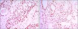
- Immunohistochemical analysis of paraffin-embedded lung cancer tissues (left) and kidney cancer tissues (right) using SIRT1 monoclonal antibody (Product # MA5-15677) followed with DAB staining.
Supportive validation
- Submitted by
- Invitrogen Antibodies (provider)
- Main image

- Experimental details
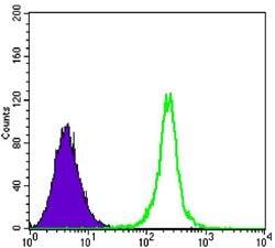
- Flow cytometric analysis of K562 cells using SIRT1 monoclonal antibody (Product # MA5-15677) (green) and negative control (purple).
- Submitted by
- Invitrogen Antibodies (provider)
- Main image

- Experimental details
- Flow cytometric analysis of K562 cells using SIRT1 monoclonal antibody (Product # MA5-15677) (green) and negative control (purple).

Supportive validation
- Submitted by
- Invitrogen Antibodies (provider)
- Main image
- Experimental details
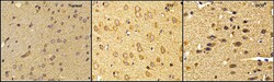
- Fig. 8 Desoxyrhapontigenin ameliorates the protein expression of Sirt-1, according to immunohistochemical analysis, in the brain tissues of rats with anaesthesia-induced neuronal injury
- Submitted by
- Invitrogen Antibodies (provider)
- Main image
- Experimental details
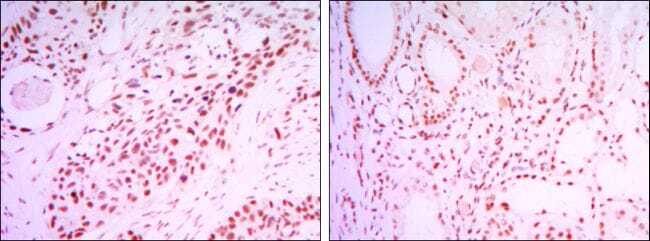
- Figure 5 In Th1-HT context, SIRT1 was downregulated while HIF-alpha was upregulated. Addition of Th1 cytokines to primary cultures of human thyrocytes induced a significant reduction of SIRT1 ( A ) and an increase of HIF-1alpha protein ( B ). Densitometric values were normalized against the beta-actin level. Results are expressed as means +- SEM from seven (SIRT1) and four (HIF-1alpha) experiments ( n = 4-7) at least in duplicate. SIRT1 protein expression ( C ) was significantly decreased in HT patients while HIF-1alpha ( D ) was increased. Densitometric values were normalized against the GAPDH level. Results are expressed as means +- SEM from five or six individual samples ( n = 5-6). * p < 0.05, *** p < 0.005 compared to controls. Representative blots are shown. Paranodular tissue from multinodular goiter patients designated as controls ( E , H ) and HT thyroid samples ( F , G , I , J ) were used to perform IHC with SIRT1 and HIF-1alpha specific antibodies. In controls ( E ), SIRT1 was detected in thyrocytes of hypofunctional follicles (+) and more intensively in active follicles (*). In HT ( F , G ), staining was less intense in altered active-like follicles ($). In control thyroids ( H ), HIF-1alpha staining was observed in the cytoplasm of the thyrocytes (+ and *). In HT thyroids ( I , J ), non-inflammatory and inflammatory zones displayed hypofunctional (+) and altered active-like ($) follicles with strong staining of HIF-1alpha in thyrocyte cytoplasm. Illustrations sho