 Explore
 Validate
 Learn
Learn Western blot
Antibody data
- Antibody Data
- Antigen structure
- References [4]
- Comments [0]
- Validations
- Western blot [3]
- Immunocytochemistry [1]
- Other assay [1]
Submit
Validation data
Reference
Comment
Report error
- Product number
- PA5-17074 - Provider product page

- Provider
- Invitrogen Antibodies
- Product name
- SIRT1 Polyclonal Antibody
- Antibody type
- Polyclonal
- Antigen
- Synthetic peptide
- Description
- It is not recommended to aliquot this antibody.
- Concentration
- 316 µg/mL
Submitted references Vitamin D Prevents High Glucose-Induced Lipid Droplets Accumulation in Cultured Endothelial Cells: The Role of Thioredoxin Interacting Protein.
Acid Loading Unmasks Glucose Homeostatic Instability in Proximal-Tubule-Targeted Insulin/Insulin-Like-Growth-Factor-1 Receptor Dual Knockout Mice.
Sirtuin1 promotes osteogenic differentiation through downregulation of peroxisome proliferator-activated receptor γ in MC3T3-E1 cells.
MiR-132 regulates osteogenic differentiation via downregulating Sirtuin1 in a peroxisome proliferator-activated receptor β/δ-dependent manner.
Scrimieri R, Cazzaniga A, Castiglioni S, Maier JAM
Biomedicines 2021 Dec 10;9(12)
Biomedicines 2021 Dec 10;9(12)
Acid Loading Unmasks Glucose Homeostatic Instability in Proximal-Tubule-Targeted Insulin/Insulin-Like-Growth-Factor-1 Receptor Dual Knockout Mice.
Aljaylani A, Fluitt M, Piselli A, Shepard BD, Tiwari S, Ecelbarger CM
Cellular physiology and biochemistry : international journal of experimental cellular physiology, biochemistry, and pharmacology 2020 Jul 18;54(4):682-695
Cellular physiology and biochemistry : international journal of experimental cellular physiology, biochemistry, and pharmacology 2020 Jul 18;54(4):682-695
Sirtuin1 promotes osteogenic differentiation through downregulation of peroxisome proliferator-activated receptor γ in MC3T3-E1 cells.
Qu B, Ma Y, Yan M, Gong K, Liang F, Deng S, Jiang K, Ma Z, Pan X
Biochemical and biophysical research communications 2016 Sep 9;478(1):439-445
Biochemical and biophysical research communications 2016 Sep 9;478(1):439-445
MiR-132 regulates osteogenic differentiation via downregulating Sirtuin1 in a peroxisome proliferator-activated receptor β/δ-dependent manner.
Gong K, Qu B, Liao D, Liu D, Wang C, Zhou J, Pan X
Biochemical and biophysical research communications 2016 Sep 9;478(1):260-267
Biochemical and biophysical research communications 2016 Sep 9;478(1):260-267
No comments: Submit comment
Supportive validation
- Submitted by
- Invitrogen Antibodies (provider)
- Main image

- Experimental details
- Western blot analysis of extracts from F9, C2C12 and PYS-2 cells using SirT1 polyclonal antibody (Product # PA5-17074).

- Submitted by
- Invitrogen Antibodies (provider)
- Main image
- Experimental details
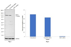
- Knockdown of SIRT1 was achieved by transfecting C2C12 with SIRT1 specific siRNAs (Silencer® select Product # S96766, S96765). Western blot analysis (Fig. a) was performed using Nuclear enriched extracts from the SIRT1 knockdown cells (lane 3), non-targeting scrambled siRNA transfected cells (lane 2) and untransfected cells (lane 1). The blot was probed with SIRT1 Polyclonal Antibody (Product # PA5-17074, 1:1000 dilution) and Goat anti-Rabbit IgG (H+L) Superclonal™ Recombinant Secondary Antibody, HRP (Product # A27036, 1:4000 dilution). Densitometric analysis of this western blot is shown in histogram (Fig. b). Decrease in signal upon siRNA mediated knock down confirms that antibody is specific to SIRT1.

- Submitted by
- Invitrogen Antibodies (provider)
- Main image
- Experimental details
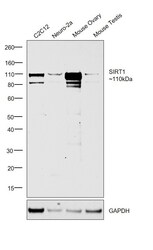
- Western blot was performed using Anti-SIRT1 Polyclonal Antibody (Product # PA5-17074) and a 110kDa band corresponding to SIRT1 was observed across all cell lines and tissues tested. Nuclear enriched extracts (50 µg lysate) of C2C12 (Lane 1), Neuro-2a (Lane 2), Mouse Ovary (Lane 3) and Mouse Testis (Lane 4) were electrophoresed using NuPAGE™ 4-12% Bis-Tris Protein Gel (Product # NP0321BOX). Resolved proteins were then transferred onto a Nitrocellulose membrane (Product # IB23001) by iBlot® 2 Dry Blotting System (Product # IB21001). The blot was probed with the primary antibody (1:1000 dilution) and detected by chemiluminescence with Goat anti-Rabbit IgG (H+L) Superclonal™ Recombinant Secondary Antibody, HRP (Product # A27036, 1:4000 dilution) using the iBright FL 1000 (Product # A32752). Chemiluminescent detection was performed using Novex® ECL Chemiluminescent Substrate Reagent Kit (Product # WP20005).

Supportive validation
- Submitted by
- Invitrogen Antibodies (provider)
- Main image

- Experimental details
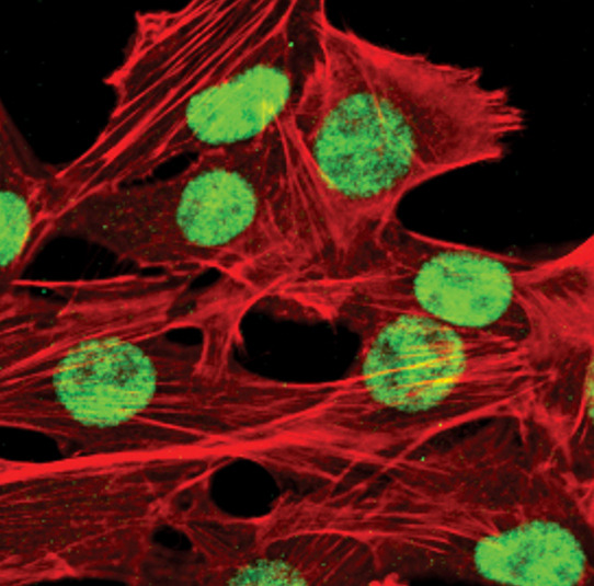
- Immunofluorescent analysis of SirT1 in C2C12 cells using a SirT1 polyclonal antibody (Product # PA5-17074) (green). Actin filaments are labeled with a fluorescent red phalloidin.
Supportive validation
- Submitted by
- Invitrogen Antibodies (provider)
- Main image

- Experimental details
- Figure 1 High glucose upregulates TXNIP and downregulates SIRT1 in HUVEC. Western blot (left panel) was performed on cell lysates using specific antibodies against TXNIP, SIRT1, HSP70, PON2, SIRT2, and SOD2. Actin was used as an equal loading control. A representative blot is shown. Densitometric analysis (right panel) was performed using Image J Lab software on three different blots, and the results are the mean of three independent experiments +- SD. * p < 0.05.
