Explore
 Validate
 Learn
Learn Western blot
 ELISA
ELISA Immunocytochemistry
Antibody data
- Antibody Data
- Antigen structure
- References [2]
- Comments [0]
- Validations
- Immunocytochemistry [4]
- Immunohistochemistry [1]
- Other assay [1]
Submit
Validation data
Reference
Comment
Report error
- Product number
- MA5-15840 - Provider product page

- Provider
- Invitrogen Antibodies
- Product name
- GRK2 Monoclonal Antibody (3F8)
- Antibody type
- Monoclonal
- Antigen
- Purifed from natural sources
- Description
- MA5-15840 targets GRK2 in indirect ELISA, IF, IHC, and WB applications and shows reactivity with Human, mouse, Non-human primate, and Rat samples. The MA5-15840 immunogen is purified recombinant fragment of human GRK2 expressed in E. Coli. . MA5-15840 detects GRK2 which has a predicted molecular weight of approximately 80kDa.
- Reactivity
- Human, Mouse, Rat
- Host
- Mouse
- Isotype
- IgG
- Antibody clone number
- 3F8
- Vial size
- 100 μL
- Concentration
- Conc. Not Determined
- Storage
- Store at 4°C short term. For long term storage, store at -20°C, avoiding freeze/thaw cycles.
Submitted references GRK2 mediates β-arrestin interactions with 5-HT(2) receptors for JC polyomavirus endocytosis.
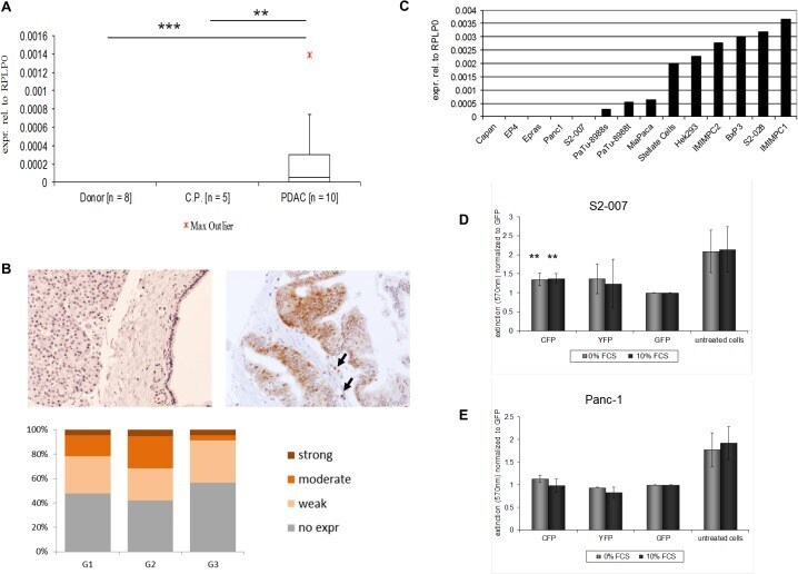
A multistep high-content screening approach to identify novel functionally relevant target genes in pancreatic cancer.
Mayberry CL, Wilczek MP, Fong TM, Nichols SL, Maginnis MS
Journal of virology 2021 Mar 10;95(7)
Journal of virology 2021 Mar 10;95(7)
A multistep high-content screening approach to identify novel functionally relevant target genes in pancreatic cancer.
Buchholz M, Honstein T, Kirchhoff S, Kreider R, Schmidt H, Sipos B, Gress TM
PloS one 2015;10(4):e0122946
PloS one 2015;10(4):e0122946
No comments: Submit comment
Supportive validation
- Submitted by
- Invitrogen Antibodies (provider)
- Main image

- Experimental details
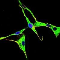
- Immunofluorescence analysis of NIH/3T3 cells using GRK2 monoclonal antibody (Product # MA5-15840) (Green). Blue: DRAQ5 fluorescent DNA dye.
- Submitted by
- Invitrogen Antibodies (provider)
- Main image

- Experimental details
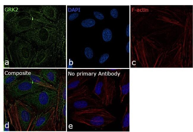
- Immunofluorescence analysis of GRK2 was performed using 70% confluent log phase HeLa cells. The cells were fixed with 4% paraformaldehyde for 10 minutes, permeabilized with 0.1% Triton™ X-100 for 15 minutes, and blocked with 1% BSA for 1 hour at room temperature. The cells were labeled with GRK2 Monoclonal Antibody (3F8) (Product # MA5-15840) at 1:250 dilution in 0.1% BSA, incubated at 4 degree Celsius overnight and then labeled with Goat anti-Mouse IgG (H+L) Superclonal™ Secondary Antibody, Alexa Fluor® 488 conjugate (Product # A28175) for 45 minutes at room temperature (Panel a: green). Nuclei (Panel b: blue) were stained with ProLong™ Diamond Antifade Mountant with DAPI (Product # P36962). F-actin (Panel c: red) was stained with Rhodamine Phalloidin (Product # R415, 1:300). Panel d represents the merged image showing cytoplasmic localization. Panel e represents control cells with no primary antibody to assess background. The images were captured at 60X magnification.

- Submitted by
- Invitrogen Antibodies (provider)
- Main image

- Experimental details
- Immunofluorescence analysis of NIH/3T3 cells using GRK2 monoclonal antibody (Product # MA5-15840) (Green). Blue: DRAQ5 fluorescent DNA dye.

- Submitted by
- Invitrogen Antibodies (provider)
- Main image

- Experimental details
- Immunofluorescence analysis of GRK2 was performed using 70% confluent log phase HeLa cells. The cells were fixed with 4% paraformaldehyde for 10 minutes, permeabilized with 0.1% Triton™ X-100 for 15 minutes, and blocked with 1% BSA for 1 hour at room temperature. The cells were labeled with GRK2 Monoclonal Antibody (3F8) (Product # MA5-15840) at 1:250 dilution in 0.1% BSA, incubated at 4 degree Celsius overnight and then labeled with Goat anti-Mouse IgG (H+L) Superclonal™ Secondary Antibody, Alexa Fluor® 488 conjugate (Product # A28175) for 45 minutes at room temperature (Panel a: green). Nuclei (Panel b: blue) were stained with ProLong™ Diamond Antifade Mountant with DAPI (Product # P36962). F-actin (Panel c: red) was stained with Rhodamine Phalloidin (Product # R415, 1:300). Panel d represents the merged image showing cytoplasmic localization. Panel e represents control cells with no primary antibody to assess background. The images were captured at 60X magnification.
Supportive validation
- Submitted by
- Invitrogen Antibodies (provider)
- Main image

- Experimental details
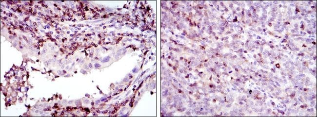
- Immunohistochemical analysis of paraffin-embedded endometrial cancer tissues (left) and cervical cancer tissues (right) using GRK2 monoclonal antibody (Product # MA5-15840) followed with DAB staining.
Supportive validation
- Submitted by
- Invitrogen Antibodies (provider)
- Main image

- Experimental details
- Fig 5 ADRBK1 overexpression promotes growth in human PDAC. A: Box-and-whisker plot showing ADRBK1 mRNA expression in primary human pancreatic tumor tissue samples, chronic pancreatitis and normal pancreas as analyzed by quantitative realtime reverse transcription PCR (qRT-PCR). Expression was normalized to ribosomal protein, large, P0 (RPLP0) mRNA levels. Data in Fig. represent median and 2nd and 3rd quartiles (boxes) as well as minimum and maximum values (whiskers). CP = Chronic Pancreatitis. ** p