Explore
 Validate
 Learn
LearnPA5-26216
antibody from Invitrogen Antibodies
Targeting: ANGPTL4
ARP4, FIAF, HFARP, NL2, PGAR, pp1158
 Western blot
 Immunohistochemistry
Antibody data
- Antibody Data
- Antigen structure
- References [2]
- Comments [0]
- Validations
- Immunohistochemistry [1]
- Flow cytometry [2]
- Other assay [2]
Submit
Validation data
Reference
Comment
Report error
- Product number
- PA5-26216 - Provider product page

- Provider
- Invitrogen Antibodies
- Product name
- ANGPTL4 Polyclonal Antibody
- Antibody type
- Polyclonal
- Antigen
- Synthetic peptide
- Reactivity
- Human
- Host
- Rabbit
- Isotype
- IgG
- Vial size
- 400 μL
- Storage
- Store at 4°C short term. For long term storage, store at -20°C, avoiding freeze/thaw cycles.
Submitted references Molecular Fingerprint for Terminal Abdominal Aortic Aneurysm Disease.
ANGPTL4 deficiency in haematopoietic cells promotes monocyte expansion and atherosclerosis progression.
Gäbel G, Northoff BH, Weinzierl I, Ludwig S, Hinterseher I, Wilfert W, Teupser D, Doderer SA, Bergert H, Schönleben F, Lindeman JHN, Holdt LM
Journal of the American Heart Association 2017 Nov 30;6(12)
Journal of the American Heart Association 2017 Nov 30;6(12)
ANGPTL4 deficiency in haematopoietic cells promotes monocyte expansion and atherosclerosis progression.
Aryal B, Rotllan N, Araldi E, Ramírez CM, He S, Chousterman BG, Fenn AM, Wanschel A, Madrigal-Matute J, Warrier N, Martín-Ventura JL, Swirski FK, Suárez Y, Fernández-Hernando C
Nature communications 2016 Jul 27;7:12313
Nature communications 2016 Jul 27;7:12313
No comments: Submit comment
Supportive validation
- Submitted by
- Invitrogen Antibodies (provider)
- Main image

- Experimental details
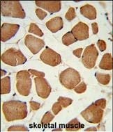
- Immunohistochemistry analysis of ANGPTL4 in formalin-fixed and paraffin-embedded human skeletal muscle. Samples were incubated with ANGPTL4 polyclonal antibody (Product # PA5-26216) which was peroxidase-conjugated to the secondary antibody, followed by DAB staining. This data demonstrates the use of this antibody for immunohistochemistry; clinical relevance has not been evaluated.
Supportive validation
- Submitted by
- Invitrogen Antibodies (provider)
- Main image
- Experimental details
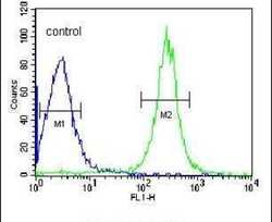
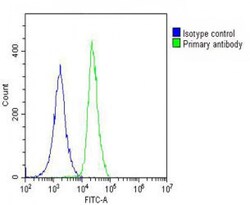
- Flow cytometry analysis of HepG2 cells using an ANGPTL4 polyclonal antibody (Product # PA5-26216) (right) compared to a negative control cell (left) at a dilution of 1:10-50, followed by a FITC-conjugated goat anti-rabbit antibody
- Submitted by
- Invitrogen Antibodies (provider)
- Main image
- Experimental details
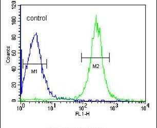
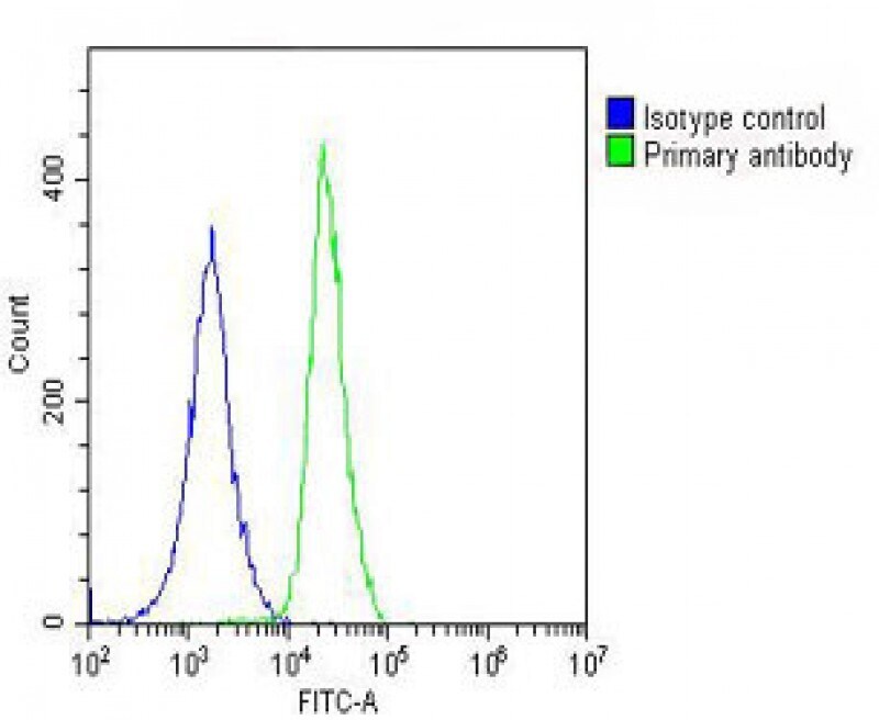
- Flow cytometry of (overlay histogram) of ANGPTL4 in A549 cells (green line). Samples were incubated with ANGPTL4 polyclonal antibody (Product # PA5-26216) using a dilution of 1:25 dilution for 60 min at 37°C followed by Goat-Anti-Rabbit IgG, DyLight® 488 Conjugated Highly Cross-Adsorbed at 1:200 dilution for 40 min at 37°C. The cells were fixed with 2% paraformaldehyde (10 min) and then permeabilized with 90% methanol for 10 min. The cells were then incubated in 2% bovine serum albumin to block non-specific protein-protein interactions followed by the primary antibody. Isotype control antibody (blue line) was rabbit IgG (1 μg/1x10^6 cells) used under the same conditions. Acquisition of >10, 000 events was performed.
Supportive validation
- Submitted by
- Invitrogen Antibodies (provider)
- Main image
- Experimental details
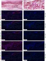
- Figure 5 Results of the immunohistochemistry. Staining for protein products of our candidate genes confirmed presence in the aneurysmatic wall. ANGPTL 4, HILPDA , SRPX 2, and LOX are localized in pericytes of microvessels in the medial-adventitial border zone. Staining for SRPX 2 is diffuse in the media and also observed in adipocytes of the adventitia. Demonstrated are representative sections from a 55-mm stable abdominal aortic aneurysm.

- Submitted by
- Invitrogen Antibodies (provider)
- Main image

- Experimental details
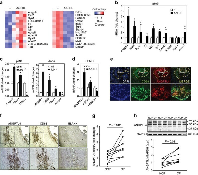
- Figure 1 ANGPTL4 is expressed in macrophages accumulated in atherosclerotic plaques. ( a ) Heat map representation of gene expression from microarray data comparing mouse peritoneal macrophages incubated with or without Ac-LDL (120 mug ml -1 ) for 24 h. Left panel shows upregulated genes ( P =1.4) and right panel shows downregulated genes ( P =2.08) upon Ac-LDL treatment compared with non-treated cells. Samples were analysed in triplicate. ( b ) qRT-PCR validation of selected genes upregulated in the microarray in mouse peritoneal macrophages treated with or without Ac-LDL for 24 h. qRT-PCR analysis of Angptl4 expression levels in macrophages ( c , left) and whole aorta ( c , right) from WT and Ldlr -/- mice fed a WD (left) ( n =4 per group), and in human peripheral blood mononuclear cells ( d ) treated with or without Ac-LDL (120 mug ml -1 ) for 24 h. Abca1 and Hmgcr genes were used as control genes for cholesterol loading and Cd68 was used as a marker for macrophages. All data represent the mean+-s.e.m. and are representative of three experiments in duplicate; * P