Explore
 Validate
 Learn
Learn Flow cytometry
Antibody data
- Antibody Data
- Antigen structure
- References [5]
- Comments [0]
- Validations
- Flow cytometry [1]
Submit
Validation data
Reference
Comment
Report error
- Product number
- 12-0039-41 - Provider product page

- Provider
- Invitrogen Antibodies
- Product name
- Anti-CD3 Monoclonal Antibody (HIT3a), PE, eBioscience™
- Antibody type
- Monoclonal
- Antigen
- Other
- Description
- Description: The HIT3a monoclonal antibody reacts with human CD3e, a 20 kDa subunit of the TCR complex. Along with the other CD3 subunits gamma and delta, the ε chain is required for proper assembly, trafficking and surface expression of the TCR complex. CD3 is expressed by thymocytes in a developmentally regulated manner and by all mature T cells. Crosslinking of TCR with HIT3a initiates an intracellular biochemical pathway resulting in cellular activation and proliferation. Applications Reported: The HIT3a antibody has been reported for use in flow cytometric analysis. Applications Tested: This HIT3a antibody has been pre-titrated and tested by flow cytometric analysis of normal human peripheral blood cells. This can be used at 5 µL (0.125 µg) per test. A test is defined as the amount (µg) of antibody that will stain a cell sample in a final volume of 100 µL. Cell number should be determined empirically but can range from 10^5 to 10^8 cells/test. Excitation: 488-561 nm; Emission: 578 nm; Laser: Blue Laser, Green Laser, Yellow-Green Laser. Filtration: 0.2 µm post-manufacturing filtered.
- Reactivity
- Human
- Host
- Mouse
- Conjugate
- Yellow dye
- Isotype
- IgG
- Antibody clone number
- HIT3a
- Vial size
- 25 Tests
- Concentration
- 5 µL/Test
- Storage
- 4° C, store in dark, DO NOT FREEZE!
Submitted references The elevated glutaminolysis of bladder cancer and T cells in a simulated tumor microenvironment contributes to the up-regulation of PD-L1 expression by interferon-γ.
A Fusion Receptor as a Safety Switch, Detection, and Purification Biomarker for Adoptive Transferred T Cells.
Blocking the recruitment of naive CD4+ T cells reverses immunosuppression in breast cancer.
Dendritic cells derived from hemozoin-loaded monocytes display a partial maturation phenotype that promotes HIV-1 trans-infection of CD4+ T cells and virus replication.
Induction of robust immune responses against human immunodeficiency virus is supported by the inherent tropism of adeno-associated virus type 5 for dendritic cells.
Wang L, Yang X, Li D, Liang Z, Chen Y, Ma G, Wang Y, Li Y, Liang Y, Niu H
OncoTargets and therapy 2018;11:7229-7243
OncoTargets and therapy 2018;11:7229-7243
A Fusion Receptor as a Safety Switch, Detection, and Purification Biomarker for Adoptive Transferred T Cells.
Wu X, Shi B, Zhang J, Shi Z, Di S, Fan M, Gao H, Wang H, Gu J, Jiang H, Li Z
Molecular therapy : the journal of the American Society of Gene Therapy 2017 Oct 4;25(10):2270-2279
Molecular therapy : the journal of the American Society of Gene Therapy 2017 Oct 4;25(10):2270-2279
Blocking the recruitment of naive CD4+ T cells reverses immunosuppression in breast cancer.
Su S, Liao J, Liu J, Huang D, He C, Chen F, Yang L, Wu W, Chen J, Lin L, Zeng Y, Ouyang N, Cui X, Yao H, Su F, Huang JD, Lieberman J, Liu Q, Song E
Cell research 2017 Apr;27(4):461-482
Cell research 2017 Apr;27(4):461-482
Dendritic cells derived from hemozoin-loaded monocytes display a partial maturation phenotype that promotes HIV-1 trans-infection of CD4+ T cells and virus replication.
Diou J, Tardif MR, Barat C, Tremblay MJ
Journal of immunology (Baltimore, Md. : 1950) 2010 Mar 15;184(6):2899-907
Journal of immunology (Baltimore, Md. : 1950) 2010 Mar 15;184(6):2899-907
Induction of robust immune responses against human immunodeficiency virus is supported by the inherent tropism of adeno-associated virus type 5 for dendritic cells.
Xin KQ, Mizukami H, Urabe M, Toda Y, Shinoda K, Yoshida A, Oomura K, Kojima Y, Ichino M, Klinman D, Ozawa K, Okuda K
Journal of virology 2006 Dec;80(24):11899-910
Journal of virology 2006 Dec;80(24):11899-910
No comments: Submit comment
Supportive validation
- Submitted by
- Invitrogen Antibodies (provider)
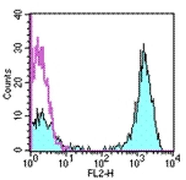
- Main image

- Experimental details
- Staining of normal human peripheral blood cells with staining buffer (autofluroescence) (open histogram) or Anti-Human CD3 PE (filled histogram). Cells in the lymphocyte gate were used for analysis.
- Conjugate
- Yellow dye