Explore
 Validate
 Learn
LearnA700-016
antibody from Invitrogen Antibodies
Targeting: CD247
CD3H, CD3Q, CD3Z
 Western blot Immunocytochemistry
 Immunoprecipitation Immunohistochemistry Flow cytometry Other assay
Antibody data
- Antibody Data
- Antigen structure
- References [0]
- Comments [0]
- Validations
- Western blot [3]
- Immunocytochemistry [1]
- Immunohistochemistry [6]
- Flow cytometry [1]
- Other assay [1]
Submit
Validation data
Reference
Comment
Report error
- Product number
- A700-016 - Provider product page

- Provider
- Invitrogen Antibodies
- Product name
- CD3E Recombinant Rabbit Monoclonal Antibody (BL-298-5D12)
- Antibody type
- Monoclonal
- Antigen
- Other
- Reactivity
- Human
- Host
- Rabbit
- Isotype
- IgG
- Antibody clone number
- BL-298-5D12
- Vial size
- 100 µL
- Concentration
- 50 µg/mL
- Storage
- 4° C
No comments: Submit comment
Supportive validation
- Submitted by
- Invitrogen Antibodies (provider)
- Main image
- Experimental details
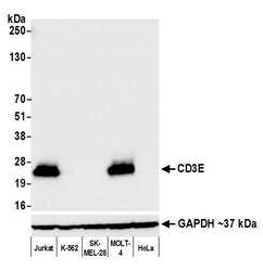
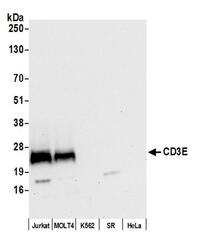
- Detection of human CD3E by western blot. Samples: Whole cell lysate (50 µg) from Jurkat, K-562, SK-MEL-28, MOLT-4, and HeLa cells prepared using NETN lysis buffer. Antibody: Rabbit anti-CD3E recombinant monoclonal antibody [BL-298-5D12] (Product # A700-016 lot 2) used at 1:1000. Secondary: HRP-conjugated goat anti-rabbit IgG (A120-101P). Chemiluminescence with an exposure time of 3 seconds. Lower Panel: Rabbit anti-GAPDH (Product # A300-639A).
- Submitted by
- Invitrogen Antibodies (provider)
- Main image
- Experimental details
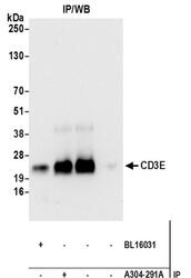
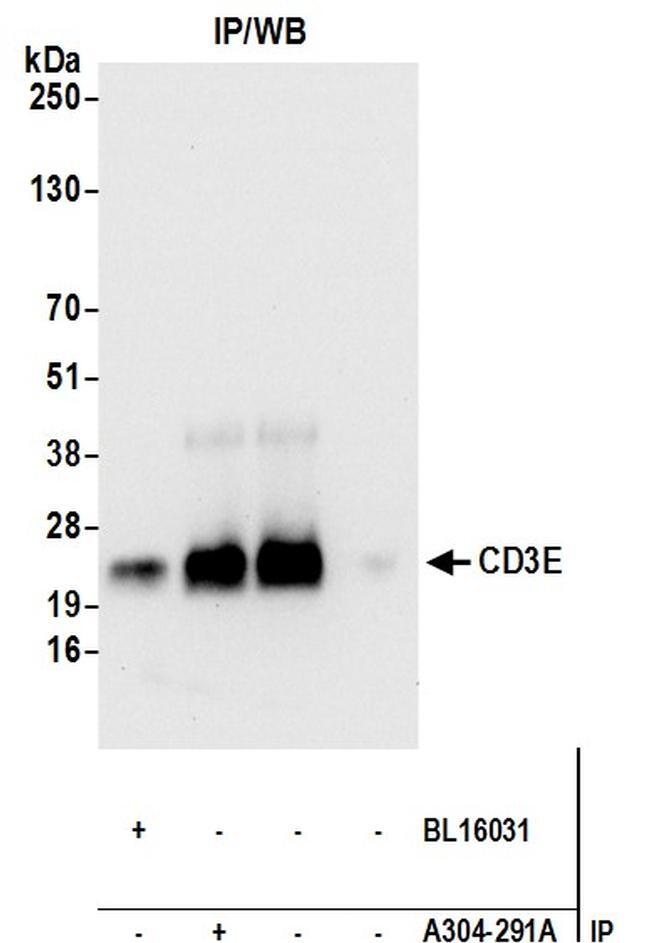
- Detection of human CD3E by WB of immunoprecipitates from Jurkat lysate. Antibodies: Rabbit anti-CD3E recombinant monoclonal [BL-298-5D12] (A700-016) and rabbit anti-CD3E antibodies (BL16031, A304-291A). Secondary: ReliaBLOT® reagents (WB120).
- Submitted by
- Invitrogen Antibodies (provider)
- Main image
- Experimental details
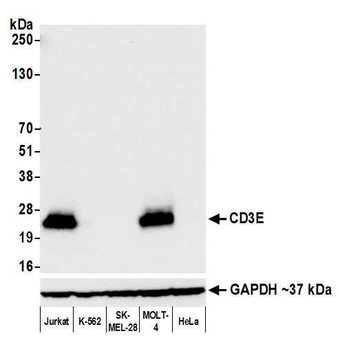
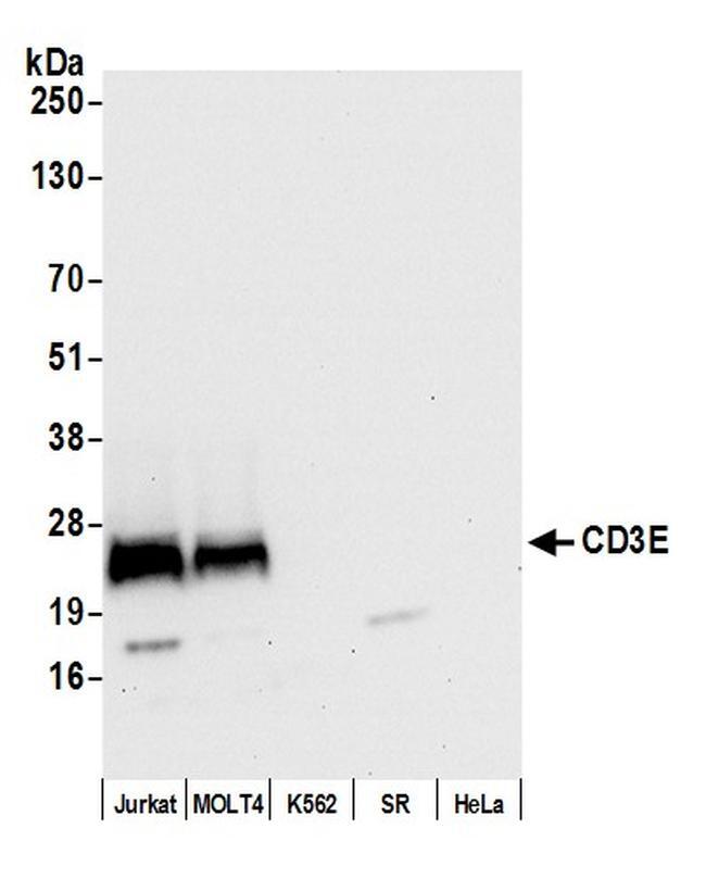
- Detection of human CD3E by WB in Jurkat, MOLT4, K562, SR, and HeLa lysate. Antibody: Rabbit anti-CD3E recombinant monoclonal [BL-298-5D12] (A700-016). Secondary: HRP-conjugated goat anti-rabbit IgG (A120-101P).
Supportive validation
- Submitted by
- Invitrogen Antibodies (provider)
- Main image
- Experimental details
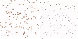
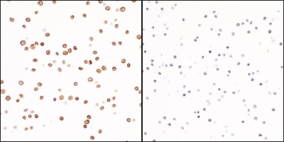
- Detection of human CD3E in FFPE Molt4 cells (left) and Ramos cells (right) by ICC. Antibody: Rabbit anti-CD3E recombinant monoclonal [BL-298-5D12] (A700-016). Secondary: HRP-conjugated goat anti-rabbit IgG (A120-501P). Substrate: DAB.
Supportive validation
- Submitted by
- Invitrogen Antibodies (provider)
- Main image
- Experimental details
- Detection of human CD3E in FFPE breast carcinoma by IHC.Antibody:Rabbit anti-CD3E recombinant monoclonal [BL-298-5D12] (Product # A700-016 lot 2).Secondary:HRP-conjugated goat anti-rabbit IgG (A120-501P).Substrate:DAB.

- Submitted by
- Invitrogen Antibodies (provider)
- Main image
- Experimental details
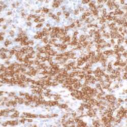
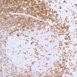
- Detection of human CD3E in FFPE tonsil by IHC.Antibody: Rabbit anti-CD3E recombinant monoclonal [BL-298-5D12] (Product # A700-016 lot 2).Secondary: HRP-conjugated goat anti-rabbit IgG (A120-501P).Substrate: DAB.

- Submitted by
- Invitrogen Antibodies (provider)
- Main image
- Experimental details
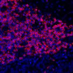
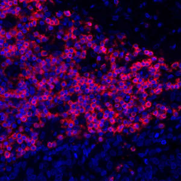
- Detection of human CD3E (red) by immunohistochemistry. Sample: FFPE section of human lung carcinoma. Antibody: Rabbit anti-CD3E recombinant monoclonal antibody [BL-298-5D12] (Product # A700-016 lot 1) used at 1:250. Secondary: HRP-conjugated goat anti-rabbit IgG (A120-501P). Substrate: Opal™. Counterstain: DAPI (blue).
- Submitted by
- Invitrogen Antibodies (provider)
- Main image
- Experimental details
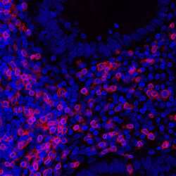
- Detection of human CD3E by immunhistochemistry.Sample: FFPE section of appendix.Antibody: Rabbit anti-CD3E recombinant monoclonal antibody [BL-298-5D12] (Product # A700-016 lot 1) used at 1:250.Secondary: DyLight® 594-conjugated goat anti-rabbit IgG (A120-101D4).
- Submitted by
- Invitrogen Antibodies (provider)
- Main image

- Experimental details
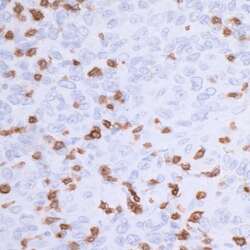
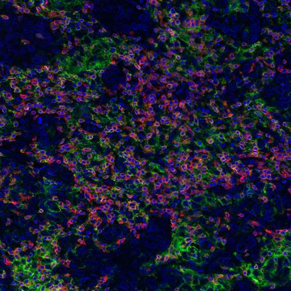
- Detection of human CD3E (orange), CD8 alpha (red), and PD-L1 (green) in FFPE breast carcinoma by IHC-IF. Rabbit anti-CD3E recombinant monoclonal [BL-298-5D12] (Product # A700-016), rabbit anti-CD8 alpha recombinant monoclonal [BLR044F] (Product # A700-044), rabbit anti-PD-L1 recombinant monoclonal [BLR020E] (Product # A700-020). Secondary: HRP-conjugated goat anti-rabbit IgG (A120-501P). Substrate: Opal™ 520, 620, and 690. Counterstain: DAPI (blue).
- Submitted by
- Invitrogen Antibodies (provider)
- Main image
- Experimental details
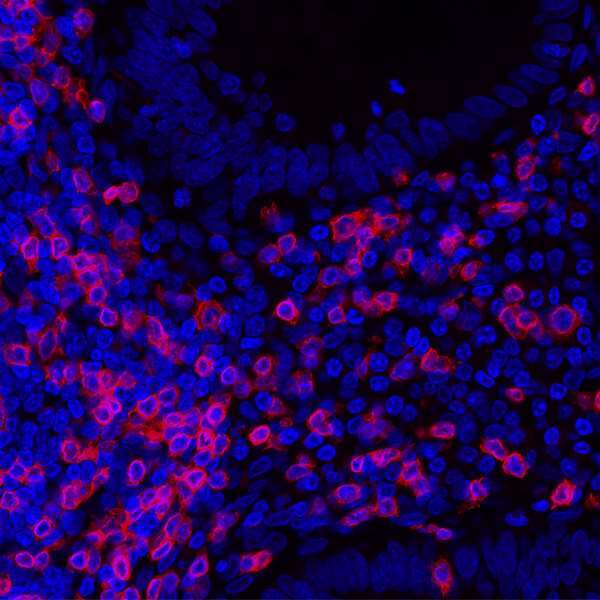
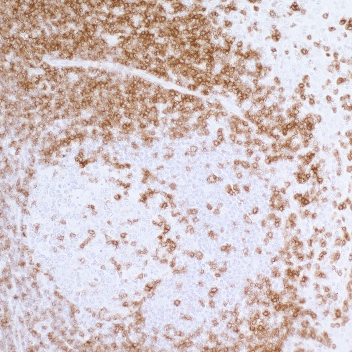
- Detection of human CD3E in FFPE tonsil by IHC. Antibody: Rabbit anti-CD3E recombinant monoclonal [BL-298-5D12] (A700-016). Secondary: HRP-conjugated goat anti-rabbit IgG (A120-501P). Substrate: DAB.
Supportive validation
- Submitted by
- Invitrogen Antibodies (provider)
- Main image
- Experimental details
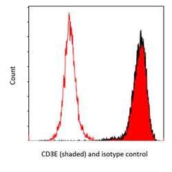
- Detection of human CD3E by flow cytometry. Sample: Jurkat cells were fixed in 4% formaldehyde and permeabilized with 90% methanol. Antibody: 1E6 cells were incubated with 1 µL of rabbit anti-CD3E recombinant monoclonal antibody [BL-298-5D12] (Product # A700-016; lot 1) (red shaded) or isotype control (unshaded). Secondary: DyLight® 650-conjugated goat anti-rabbit IgG (Product # A120-201D5).

Supportive validation
- Submitted by
- Invitrogen Antibodies (provider)
- Main image
- Experimental details
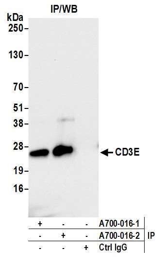
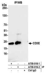
- Detection of human CD3E by western blot of immunoprecipitates. Samples: Whole cell lysate (1.0 mg per IP reaction; 20% of IP loaded) from Jurkat cells prepared using NETN lysis buffer. Antibodies: Rabbit anti-CD3E recombinant monoclonal antibody [BL-298-5D12] (Product # A700-016 lot 2) used for IP at 20 µL/mg lysate. CD3E was also immunoprecipitated by a previous lot of this antibody (lot A700-016-1). For blotting immunoprecipitated CD3E (Product # A700-016) was used at 1:1000. Chemiluminescence with an exposure time of 3 seconds.