Explore
 Validate
 Learn
Learn Western blot
Antibody data
- Antibody Data
- Antigen structure
- References [0]
- Comments [0]
- Validations
- Western blot [1]
- Immunohistochemistry [1]
- Flow cytometry [2]
Submit
Validation data
Reference
Comment
Report error
- Product number
- STJ11101284 - Provider product page

- Provider
- St John's Laboratory
- Product name
- Anti-CD247 antibody [ARC0533] (STJ11101284)
- Antibody type
- Monoclonal
- Description
- Rabbit monoclonal antibody anti-CD3 zeta is suitable for use in Western Blot, Immunohistochemistry and Flow Cytometry.
- Reactivity
- Human
- Host
- Rabbit
- Conjugate
- Unconjugated
- Antigen sequence
NA- Epitope
- NA
- Isotype
- IgG
- Antibody clone number
- NA
- Vial size
- NA
- Concentration
- NA
- Storage
- Store in a freezer at-20°C and avoid freeze-thaw cycles.
- Handling
- NA
No comments: Submit comment
Supportive validation
- Submitted by
- St John's Laboratory (provider)
- Main image
- Experimental details
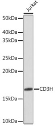
- Western blot analysis of extracts of Jurkat cells, using CD3H rabbit monoclonal antibody (STJ11101284) at 1:1000 dilution. Secondary antibody: HRP Goat Anti-rabbit IgG (H+L) at 1:10000 dilution. Lysates/proteins: 25ug per lane. Blocking buffer: 3% nonfat dry milk in TBST. Detection: ECL Enhanced Kit. Exposure time: 3min.
- Sample type
- NA
- Validation comment
- NA
- Primary Ab dilution
- NA
- Other comments
- NA
- Secondary Ab
- NA
- Secondary Ab dilution
- NA
- Protocol
- NA

Supportive validation
- Submitted by
- St John's Laboratory (provider)
- Main image
- Experimental details
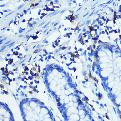
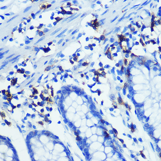
- Immunohistochemistry of paraffin-embedded human colon using CD3H rabbit monoclonal antibody (STJ11101284) at dilution of 1:100 (40x lens). Perform microwave antigen retrieval with 10 mM PBS buffer pH 7. 2 before commencing with immunohistochemistry staining protocol.
- Sample type
- NA
- Validation comment
- NA
- Primary Ab dilution
- NA
- Other comments
- NA
- Secondary Ab
- NA
- Secondary Ab dilution
- NA
- Protocol
- NA
Supportive validation
Supportive validation
- Submitted by
- St John's Laboratory (provider)
- Main image
- Experimental details
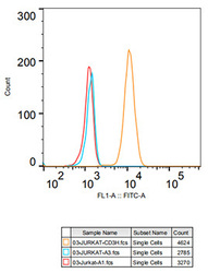
- Flow cytometry: Jurkat cells were stained with rabbit IgG isotype control (AC042, 10 ug/ml, blue line) or CD3H rabbit monoclonal antibody (STJ11101284, 10 ug/ml orange line) , followed by goat anti-rabbit polyclonal antibody FITC (1:200 dilution) staining. Non-fluorescently stained Jurkat cells was used as blank control (red line).
- Sample type
- NA
- Validation comment
- NA
- Primary Ab dilution
- NA
- Other comments
- NA
- Secondary Ab
- NA
- Secondary Ab dilution
- NA
- Protocol
- NA

Supportive validation
- Submitted by
- St John's Laboratory (provider)
- Main image
- Experimental details
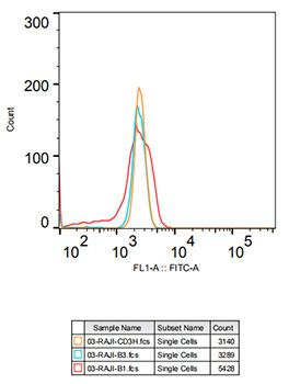
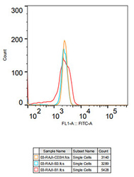
- Flow cytometry: Raji cells were stained with rabbit IgG isotype control (AC042, 10 ug/ml, blue line) or CD3H rabbit monoclonal antibody (STJ11101284, 2. 5 ug/ml orange line) , followed by goat anti-rabbit polyclonal antibody FITC (1:200 dilution) staining. Non-fluorescently stained Raji cells was used as blank control (red line).
- Sample type
- NA
- Validation comment
- NA
- Primary Ab dilution
- NA
- Other comments
- NA
- Secondary Ab
- NA
- Secondary Ab dilution
- NA
- Protocol
- NA