 Explore
 Validate
 Learn
Learn Western blot
Antibody data
- Antibody Data
- Antigen structure
- References [1]
- Comments [0]
- Validations
- Western blot [1]
- Immunocytochemistry [3]
- Immunohistochemistry [2]
- Flow cytometry [1]
- Other assay [1]
Submit
Validation data
Reference
Comment
Report error
- Product number
- MA5-32787 - Provider product page

- Provider
- Invitrogen Antibodies
- Product name
- TLR2 Recombinant Rabbit Monoclonal Antibody (JM22-41)
- Antibody type
- Monoclonal
- Antigen
- Recombinant full-length protein
- Description
- Recombinant rabbit monoclonal antibodies are produced using in vitro expression systems. The expression systems are developed by cloning in the specific antibody DNA sequences from immunoreactive rabbits. Then, individual clones are screened to select the best candidates for production. The advantages of using recombinant rabbit monoclonal antibodies include: better specificity and sensitivity, lot-to-lot consistency, animal origin-free formulations, and broader immunoreactivity to diverse targets due to larger rabbit immune repertoire.
- Reactivity
- Human, Mouse, Rat
- Host
- Rabbit
- Isotype
- IgG
- Antibody clone number
- JM22-41
- Vial size
- 100 µL
- Concentration
- 1 mg/mL
- Storage
- Store at 4°C short term. For long term storage, store at -20°C, avoiding freeze/thaw cycles.
Submitted references hTERT-immortalized gingival fibroblasts respond to cytokines but fail to mimic primary cell responses to Porphyromonas gingivalis.
Lagosz-Cwik KB, Wielento A, Lipska W, Kantorowicz M, Darczuk D, Kaczmarzyk T, Gibbs S, Potempa J, Grabiec AM
Scientific reports 2021 May 24;11(1):10770
Scientific reports 2021 May 24;11(1):10770
No comments: Submit comment
Supportive validation
- Submitted by
- Invitrogen Antibodies (provider)
- Main image
- Experimental details
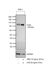
- Western blot was performed using Anti-TLR2 Rabbit Monoclonal Antibody (Product # MA5-32787) and a 90 kDa band corresponding to TLR2 was observed in cell line tested and increased upon treatment in THP-1. Membrane enriched extracts (30 µg lysate) of THP-1 (Lane 1) and THP-1 treated with PMA (10 ng/ml for 20 hours) and LPS (20 ng/ml for 6 hours) (Lane 2) were electrophoresed using Novex® NuPAGE® 4-12% Bis-Tris Protein Gel (Product # NP0322BOX). Resolved proteins were then transferred onto a nitrocellulose membrane (Product # IB23001) by iBlot® 2 Dry Blotting System (Product # IB21001). The blot was probed with the primary antibody (1:1000 dilution) and detected by chemiluminescence with Goat anti-Rabbit IgG (H+L) Superclonal™ Recombinant Secondary Antibody, HRP (Product # A27036, 1:4000 dilution) using the iBright FL 1000 (Product # A32752). Chemiluminescent detection was performed using Novex® ECL Chemiluminescent Substrate Reagent Kit (Product # WP20005).

Supportive validation
- Submitted by
- Invitrogen Antibodies (provider)
- Main image

- Experimental details
- Immunocytochemical analysis of TLR2 in PC-3M cells using a TLR2 Monoclonal antibody (Product # MA5-32787) as seen in green. The nuclear counter stain is DAPI (blue). Cells were fixed in paraformaldehyde, permeabilised with 0.25% Triton X100/PBS.

- Submitted by
- Invitrogen Antibodies (provider)
- Main image

- Experimental details
- Immunocytochemical analysis of TLR2 in A549 cells using a TLR2 Monoclonal antibody (Product # MA5-32787) as seen in green. The nuclear counter stain is DAPI (blue). Cells were fixed in paraformaldehyde, permeabilised with 0.25% Triton X100/PBS.

- Submitted by
- Invitrogen Antibodies (provider)
- Main image

- Experimental details
- Immunocytochemical analysis of TLR2 in HepG2 cells using a TLR2 Monoclonal antibody (Product # MA5-32787) as seen in green. The nuclear counter stain is DAPI (blue). Cells were fixed in paraformaldehyde, permeabilised with 0.25% Triton X100/PBS.

Supportive validation
- Submitted by
- Invitrogen Antibodies (provider)
- Main image

- Experimental details
- Immunohistochemical analysis of TLR2 of paraffin-embedded Mouse spleen tissue using a TLR2 Monoclonal antibody (Product #MA5-32787). Counter stained with hematoxylin.

- Submitted by
- Invitrogen Antibodies (provider)
- Main image

- Experimental details
- Immunohistochemical analysis of TLR2 of paraffin-embedded Human spleen tissue using a TLR2 Monoclonal antibody (Product #MA5-32787). Counter stained with hematoxylin.

Supportive validation
- Submitted by
- Invitrogen Antibodies (provider)
- Main image

- Experimental details
- Flow Cytometric analysis of TLR2 in THP-1 cells using a TLR2 Monoclonal Antibody (Product # MA5-32787) at a dilution of 1:100, as seen in red compared with an unlabelled control (cells without incubation with primary antibody; black).

Supportive validation
- Submitted by
- Invitrogen Antibodies (provider)
- Main image

- Experimental details
- Figure 4 TIGFs lack TLR2 expression and fail to respond to TLR2 agonists. ( a ) Western blot analysis of TLR2 protein expression in PHGF and TIGF total cell lysates. U-251 MG cells overexpressing hTLR2 were used as a positive control and actin was used as a loading control. Results presented are representative of two independent experiments and full-length blots are presented in Supplementary Fig. S2 . ( b ) Relative mRNA expression of TLR2 in PHGFs and TIGFs (n = 3) that were left unstimulated (med) or were stimulated with TNFalpha (10 ng/ml) or IL-1beta (10 ng/ml) for 24 h. ( c ) Relative mRNA expression of IL6 , IL8 and COX2 in PHGFs and TIGFs stimulated with Pam2CSK4 (1 mug/ml) or Pam3CSK4 (1 mug/ml) for 24 h. Data for PHGFs from individual donors (n = 3) and TIGFs from independent experiments (n = 3) are shown on a heat map as column Z-scores calculated from DeltaCt values relative to a housekeeping gene ( RPLP0 ). ( d ) Secretion of IL-6 and IL-8 by PHGFs and TIGFs (n = 3) and stimulated as in ( c ). ( e ) TLR2 promoter methylation in PHGFs and TIGFs (n = 3) analyzed using the EpiTect methyl II PCR assay and presented as mean percentage of methylated DNA + SEM. ( f ) Relative TLR4 mRNA expression in PHGFs and TIGFs (n = 3). ( g ) Western blot analysis of TLR4 protein expression in total cell lysates of PHGFs and TIGFs. Actin was used as a loading control. ( h ) Secretion of IL-6 and IL-8 by PHGFs and TIGFs (n = 3-4) infected with increasing MOI of F. nucleatum (10, 50)
