Explore
 Validate
 Learn
Learn Immunocytochemistry
 Immunoprecipitation
Antibody data
- Antibody Data
- Antigen structure
- References [3]
- Comments [0]
- Validations
- Immunocytochemistry [1]
- Flow cytometry [1]
Submit
Validation data
Reference
Comment
Report error
- Product number
- 14-9022-82 - Provider product page

- Provider
- Invitrogen Antibodies
- Product name
- Anti-CD282 (TLR2) Monoclonal Antibody (mT2.7), eBioscience™
- Antibody type
- Monoclonal
- Antigen
- Other
- Description
- Description: The mT2.7 monoclonal antibody reacts with mouse Toll-like receptor 2 (TLR2). Mouse TLR2 is expressed by the myeloid lineage, including macrophage and dendritic cells in splenocytes and the RAW264.7 cell line. To date, at least twelve members of the Toll family have been identified in human and mouse. This family of type I transmembrane protein is characterized by an extracellular domain with leucine-rich repeats and a cytoplasmic domain with homology to the type I IL-1 receptor. Two of these receptors, TLR2 and TLR4, are pattern recognition receptors and signaling molecules in response to bacterial lipoproteins and have been implicated in innate immunity and inflammation. TLR2 is expressed on the surface of cells and is responsible for distinguishing different pathogens. Applications Reported: This mT2.7 antibody has been reported for use in flow cytometric analysis, immunoprecipitation, and immunohistochemical staining of frozen tissue sections. Applications Tested: The mT2.7 antibody has been tested by flow cytometric analysis of Raw246.7 cells. This can be used at less than or equal to 0.5 µg per test. A test is defined as the amount (µg) of antibody that will stain a cell sample in a final volume of 100 µL. Cell number should be determined empirically but can range from 10^5 to 10^8 cells/test. It is recommended that the antibody be carefully titrated for optimal performance in the assay of interest. Purity: Greater than 90%, as determined by SDS-PAGE. Aggregation: Less than 10%, as determined by HPLC. Filtration: 0.2 µm post-manufacturing filtered.
- Reactivity
- Human, Mouse
- Host
- Mouse
- Isotype
- IgG
- Antibody clone number
- mT2.7
- Vial size
- 100 µg
- Concentration
- 0.5 mg/mL
- Storage
- 4° C
Submitted references Immunomodulatory activity of a novel, synthetic beta-glucan (β-glu6) in murine macrophages and human peripheral blood mononuclear cells.
TLR4 and NKT cell synergy in immunotherapy against visceral leishmaniasis.
Yersinia pestis and host macrophages: immunodeficiency of mouse macrophages induced by YscW.
Li X, Wang J, Wang W, Liu C, Sun S, Gu J, Wang X, Boraschi D, Huang Y, Qu D
PloS one 2013;8(11):e80399
PloS one 2013;8(11):e80399
TLR4 and NKT cell synergy in immunotherapy against visceral leishmaniasis.
Karmakar S, Bhaumik SK, Paul J, De T
PLoS pathogens 2012;8(4):e1002646
PLoS pathogens 2012;8(4):e1002646
Yersinia pestis and host macrophages: immunodeficiency of mouse macrophages induced by YscW.
Bi Y, Du Z, Han Y, Guo Z, Tan Y, Zhu Z, Yang R
Immunology 2009 Sep;128(1 Suppl):e406-17
Immunology 2009 Sep;128(1 Suppl):e406-17
No comments: Submit comment
Supportive validation
- Submitted by
- Invitrogen Antibodies (provider)
- Main image

- Experimental details
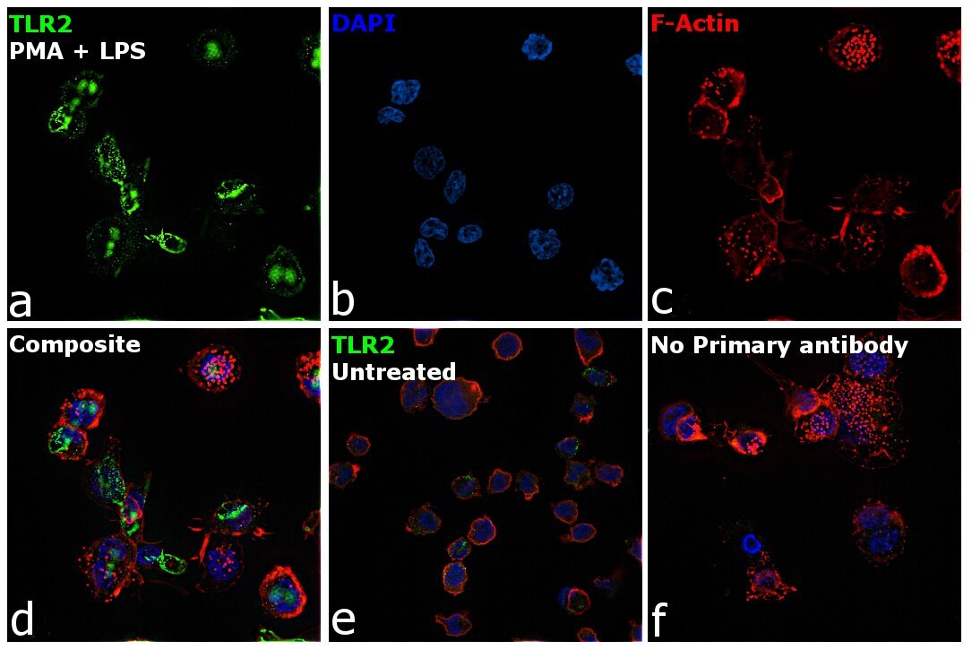
- Immunofluorescence analysis of TLR2 was performed using THP-1 cells and THP-1 treated with PMA (10 ng/mL, 20 hours) and LPS (20 ng/mL, 6 hours). The cells were fixed with 4% paraformaldehyde for 10 minutes and blocked with 2% BSA for 1 hour at room temperature. The cells were labeled with TLR2 Mouse Monoclonal Antibody (Product # 14-9022-82) at 1:100 dilution in 0.1% BSA and incubated overnight at 4 degree and then labeled with Goat anti-Mouse IgG (H+L) Highly Cross-Adsorbed Secondary Antibody, Alexa Fluor Plus 488 (Product # A32723) at a dilution of 1:2000 for 45 minutes at room temperature (Panel a: green) in THP-1 treated cells. Nuclei (Panel b: blue) were stained with ProLong™ Diamond Antifade Mountant with DAPI (Product # P36962). F-actin (Panel c: red) was stained with Rhodamine Phalloidin (Product # R415, 1:300). Panel d represents the merged image of THP-1 treated cells, which shows higher expression for TLR2 protein showing localization in lipid rafts in membrane. Panel e represents the merged image of THP-1 cells, that shows lower expression for TLR2 protein. Panel f represents control cells with no primary antibody to assess background. The images were captured at 60X magnification.
Supportive validation
- Submitted by
- Invitrogen Antibodies (provider)
- Main image

- Experimental details
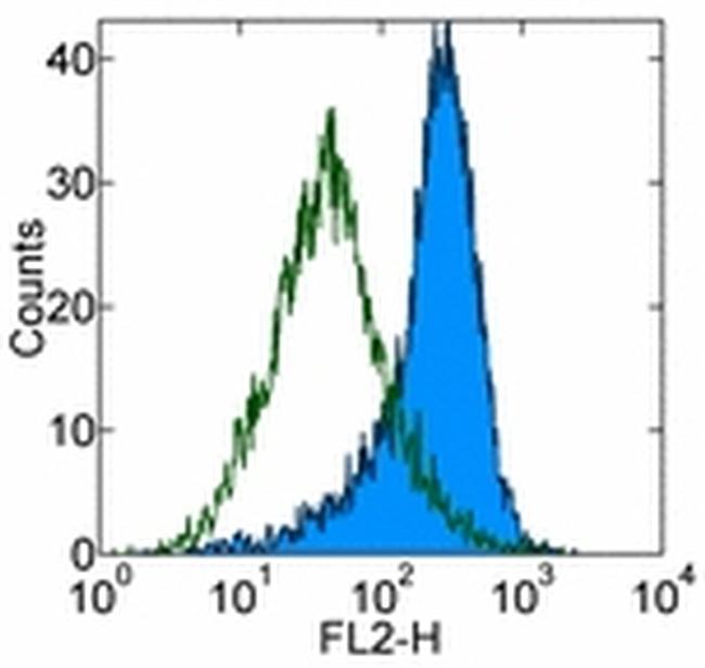
- Staining of Raw246-7 cells with 0.25 µg of Mouse IgG2a K Isotype Control Purified (Product # 14-4724-82) (open histogram) or 0.25 µg of Anti-Mouse CD282 (TLR2) Purified (filled histogram) followed by Anti-Mouse IgG Biotin (Product # 13-4013-85) and Streptavidin PE (Product # 12-4317-87).Total viable cells were used for analysis.