Explore
 Validate
 Learn
Learn Western blot
 Immunocytochemistry
Antibody data
- Antibody Data
- Antigen structure
- References [1]
- Comments [0]
- Validations
- Immunocytochemistry [1]
- Immunohistochemistry [2]
- Other assay [1]
Submit
Validation data
Reference
Comment
Report error
- Product number
- PA5-34554 - Provider product page

- Provider
- Invitrogen Antibodies
- Product name
- TLX2 Polyclonal Antibody
- Antibody type
- Polyclonal
- Antigen
- Synthetic peptide
- Description
- A suggested positive control is rat brain tissue lysate. PA5-34554 can be used with blocking peptide PEP-1597.
- Reactivity
- Human, Mouse, Rat
- Host
- Rabbit
- Isotype
- IgG
- Vial size
- 100 μg
- Concentration
- 1 mg/mL
- Storage
- Maintain refrigerated at 2-8°C for up to 3 months. For long term storage store at -20°C
Submitted references Essential roles of exosome and circRNA_101093 on ferroptosis desensitization in lung adenocarcinoma.
Zhang X, Xu Y, Ma L, Yu K, Niu Y, Xu X, Shi Y, Guo S, Xue X, Wang Y, Qiu S, Cui J, Wang H, Tian X, Miao Y, Meng F, Qiao Y, Yu Y, Wang J
Cancer communications (London, England) 2022 Apr;42(4):287-313
Cancer communications (London, England) 2022 Apr;42(4):287-313
No comments: Submit comment
Supportive validation
- Submitted by
- Invitrogen Antibodies (provider)
- Main image
- Experimental details
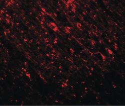
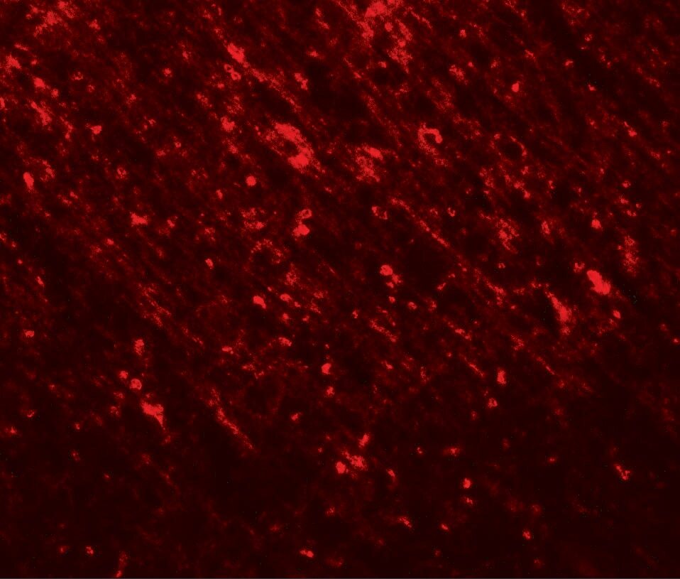
- Immunofluorescent analysis of rat brain tissue using a TLX2 polyclonal antibody (Product # PA5-34554) at a 20 µg/mL dilution.
Supportive validation
- Submitted by
- Invitrogen Antibodies (provider)
- Main image

- Experimental details
- Immunohistochemistry of TLX2 in rat brain tissue with TLX2 Polyclonal Antibody (Product # PA5-34554) at 2.5 µg/mL.

- Submitted by
- Invitrogen Antibodies (provider)
- Main image
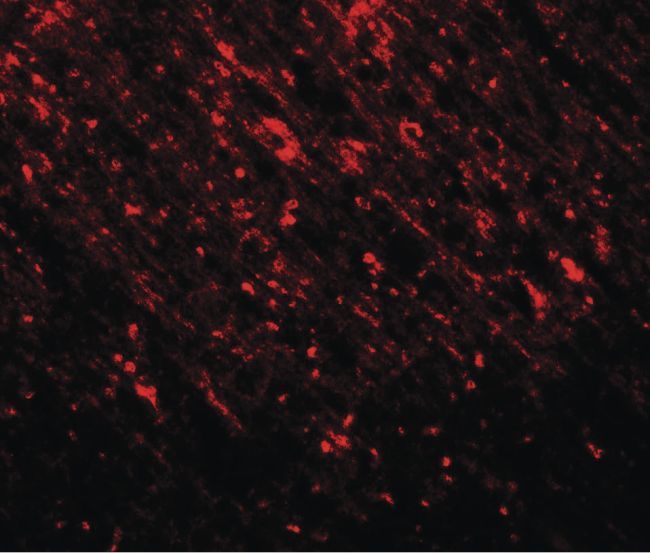
- Experimental details
- Immunofluorescence of TLX2 in rat brain tissue with TLX2 Polyclonal Antibody (Product # PA5-34554) at 20 µg/mL.
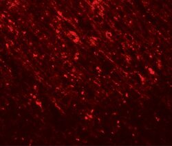
Supportive validation
- Submitted by
- Invitrogen Antibodies (provider)
- Main image
- Experimental details
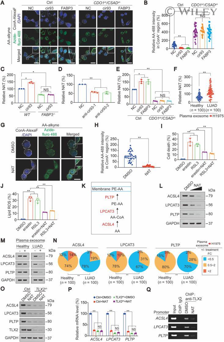
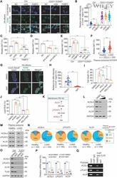
- 6 FIGURE NAT suppressed AA incorporation into the plasma membrane. (A-B) AA incorporation into the plasma membrane, as measured by a click chemistry-based method in control and H1975 cells infected with shRNAs targeting against CDO1 and CSAD followed by treatment with or without cir93 or FABP3 overexpression plasmids. Representative images are shown in panel A, and data were graphed in panel B. Scale bar, 50 mum. (C) NAT was measured in WT and FABP3 -/- H1975 cells with or without cir93 overexpression. (D) NAT in control and A549 cells treated with indicated anti-cir93. (E) NAT in control cells and H1975 cells infected with shRNAs targeting against CDO1 and CSAD followed by treatment with or without cir93 or FABP3 overexpression plasmids. (F) NAT in H1975 cells following co-incubation with plasma exosome from healthy individuals ( n = 100) and LUAD patients ( n = 100). The relevant information of healthy individual and LUAD patient is summarized in Supplementary Table S7. (G-H) Incorporation of AA into the plasma membrane in H1975 cells treated with DMSO or NAT (20 mumol/L, 24 h), as measured by a click chemistry-based method. Representative images are shown in panel G, and data were graphed in panel H. Scale bar, 20 mum. (I-J) Cell death (I) and lipid ROS generation (J) were measured in control cells and H1975 cells treated with erastin (10 mumol/L, 16-24 h) or RSL3 (5 mumol/L, 16-24 h) alone or in combination with NAT (20 mumol/L, 16-24 h). (K) Schematic presentation of how