Explore
 Validate
 Learn
Learn Western blot
Antibody data
- Antibody Data
- Antigen structure
- References [0]
- Comments [0]
- Validations
- Western blot [2]
- Immunocytochemistry [1]
- Immunohistochemistry [1]
- Other assay [3]
Submit
Validation data
Reference
Comment
Report error
- Product number
- MA5-12969 - Provider product page

- Provider
- Invitrogen Antibodies
- Product name
- S100 Monoclonal Antibody (4C4.9)
- Antibody type
- Monoclonal
- Antigen
- Purifed from natural sources
- Description
- MA5-12969 targets S100 Protein in IHC (P), ICC/IF and WB applications and shows reactivity with Bovine, Human, mouse, and Rat samples. The MA5-12969 immunogen is purified bovine brain S100 protein.
- Reactivity
- Human, Mouse, Bovine
- Host
- Mouse
- Isotype
- IgG
- Antibody clone number
- 4C4.9
- Vial size
- 500 µL
- Concentration
- 0.2 mg/mL
- Storage
- 4° C
No comments: Submit comment
Supportive validation
- Submitted by
- Invitrogen Antibodies (provider)
- Main image
- Experimental details
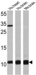
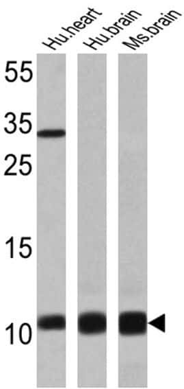
- Western blot analysis of S100 Protein was performed by loading 25 µg of human heart (lane 1), human brain (lane 2) and mouse brain (lane 3) onto an SDS polyacrylamide gel. Proteins were transferred to a PVDF membrane and blocked at 4ºC overnight. The membrane was probed with a S100 Protein monoclonal antibody (Product # MA5-12969) at a dilution of 1:200 overnight at 4°C, washed in TBST, and probed with an HRP-conjugated secondary antibody for 1 hr at room temperature in the dark. Chemiluminescent detection was performed using Pierce ECL Plus Western Blotting Substrate (Product # 32132). Results show a band at ~10 kDa.
- Submitted by
- Invitrogen Antibodies (provider)
- Main image
- Experimental details
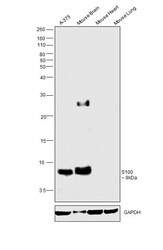
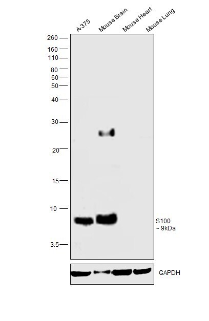
- Western blot was performed using Anti-S100 Monoclonal Antibody (Product # MA5-12969) and a 9kDa band corresponding to S100 was observed across all the cell line and tissues tested. Whole cell extracts (30 µg lysate) of A-375 (Lane 1) and tissue extracts of Mouse Brain (Lane 2), Mouse Heart (Lane 3) and Mouse Lung (Lane 4) were electrophoresed using NuPAGE™ 10% Bis-Tris Protein Gel (Product # NP0301BOX). Expression of S100 was observed to be high in Mouse brain as compared to low or negative in Mouse Heart and Mouse Lung. Resolved proteins were then transferred onto a nitrocellulose membrane (Product # IB23001) by iBlot® 2 Dry Blotting System (Product # IB21001). The blot was probed with the primary antibody (1:500 dilution) and detected by chemiluminescence with Goat anti-Mouse IgG (H+L) Superclonal™ Secondary Antibody, HRP (Product # A28177, 1:4000 dilution) using the iBright FL 1000 (Product # A32752). Chemiluminescent detection was performed using Novex® ECL Chemiluminescent Substrate Reagent Kit (Product # WP20005).
Supportive validation
- Submitted by
- Invitrogen Antibodies (provider)
- Main image
- Experimental details
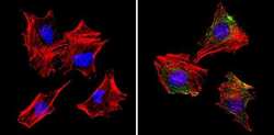
- Immunofluorescent analysis of S100 Protein (green) showing staining in the cytoplasm of C2C12 cells (right) compared to a negative control without primary antibody (left). Formalin-fixed cells were permeabilized with 0.1% Triton X-100 in TBS for 5-10 minutes and blocked with 3% BSA-PBS for 30 minutes at room temperature. Cells were probed with a S100 Protein monoclonal antibody (Product # MA5-12969) in 3% BSA-PBS at a dilution of 1:50 and incubated overnight at 4ºC in a humidified chamber. Cells were washed with PBST and incubated with a DyLight-conjugated secondary antibody in PBS at room temperature in the dark. Actin was stained using Alexa Fluor 554 (red) and nuclei were stained with Hoechst or DAPI (blue). Images were taken at a magnification of 60x.
Supportive validation
- Submitted by
- Invitrogen Antibodies (provider)
- Main image
- Experimental details
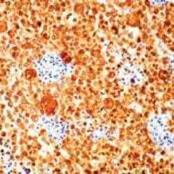
- Formalin-fixed, paraffin-embedded human melanoma stained with S-100 antibody using peroxidase-conjugate and DAB chromogen. Note cytoplasmic staining of tumor cells.

Supportive validation
- Submitted by
- Invitrogen Antibodies (provider)
- Main image
- Experimental details
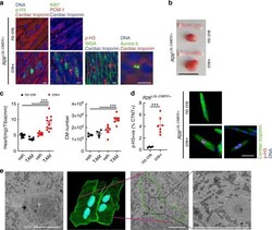
- Fig. 7 The juvenile heart is permissive to Myc-driven proliferation. a Immunofluorescent staining of cardiac troponin, p-H3, Ki67, PCM1, wheat germ agglutinin and aurora B-positive mid-body in the heart of 15-day-old control (no cre) and Myh6-Cre ; R26 LSL-CMER/+ (cre+) mice. Representative images based on analysis of five independent mice. Scale bar represents 10 mum. b Image of the whole heart and a tibia isolated from a 15-day-old control (no cre) and Myh6-Cre ; R26 LSL-CMER/+ (cre+) mice. Scale bar represents 10 mm. Representative images based on analysis on 21 independent mice. c The weight (mg) of hearts (left, R26 LSL-CMER/+ ; no cre, vehicle = 9, no cre tamoxifen = 11; R26 LSL-CMER/+ ; cre+ vehicle n = 7, cre+ tamoxifen n = 10) and quantification of the number of cardiomyocytes (right) isolated from 15-day-old control (no cre, vehicle = 3, no cre tamoxifen = 4) and Myh6-Cre ; R26 LSL-CMER/+ (cre+ vehicle n = 6, cre+ tamoxifen n = 7) mice post administration of tamoxifen or oil (veh), expressed as fold change over the length (mm) of a tibia isolated from the same mouse. Mean and s.e.m. shown. One-way ANOVA with Tukey''s multiple comparisons test; vehicle cre vs tamoxifen cre+, vehicle no cre vs tamoxifen cre+, tamoxifen no cre vs tamoxifen cre+ *** P = 0.001 (weight). Vehicle cre vs tamoxifen cre+ ** P = 0.01, vehicle no cre vs tamoxifen cre+, tamoxifen no cre vs tamoxifen cre+ *** P = 0.001 (cardiomyocyte number). Replicate samples are derived from independent mice. d

- Submitted by
- Invitrogen Antibodies (provider)
- Main image
- Experimental details
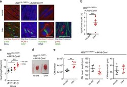
- Fig. 9 Overexpression of Cyclin T1 in the adult heart facilitates Myc-driven proliferation of adult cardiomyocytes. a Immunofluorescent staining of cardiac troponin, p-H3,Ki67, PCM1, wheat germ agglutinin (WGA) and aurora B in control ( R26 LSL-CMER/+ ; no cre) and Myh6-Cre ; R26 LSL-CMER/+ ( R26 LSL-CMER/+ ; cre+) adult mouse heart isolated 4 weeks post systemic infection with an adeno-associated virus encoding Ccnt1 ( AAV9-Ccnt1 ) and 48 h post administration of tamoxifen. Representative images based on analysis of five independent mice. Scale bars represents 10 mum (aurora B) and 50 mum (all others). b Quantification of p-H3-positive nuclei percentage in control ( R26 LSL-CMER/+ ; no cre, n = 9) and Myh6-Cre ; R26 LSL-CMER/+ ( R26 LSL-CMER/+ ; cre+, n = 7) adult mouse heart isolated 4 weeks post systemic infection with an adeno-associated virus encoding Ccnt1 ( AAV9-Ccnt1 ) and 48 h post administration of tamoxifen. Means are taken from five images per mouse; Mean and s.e.m shown. Unpaired t -test; no cre vs cre+ P < 0.0001. c The weight (mg) of hearts isolated from control ( R26 LSL-CMER/+ ; no cre, n = 9 48 h, n = 5 72 h) and Myh6-Cre ; R26 LSL-CMER/+ ( R26 LSL-CMER/+ ; cre+, n = 7 48 h, n = 6 72 h) adult mouse heart 4 weeks post systemic infection with an adeno-associated virus encoding Ccnt1 ( AAV9-Ccnt1 ) and 48 and 72 h post administration of tamoxifen, expressed as fold change over the length (mm) of a tibia isolated from the same mouse. Mean and s.e.m shown. One-wa

- Submitted by
- Invitrogen Antibodies (provider)
- Main image

- Experimental details
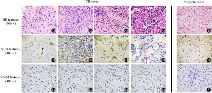
- Fig. 2 Clinical-histological features and H3K36M IHC results of CB cases and suspected case. ( A1-A4 ) The HE features of CB cases. ( B ) The HE features of the suspected case. ( C1-C4 ) The S100 features of the corresponding CB cases. ( D ) The S100 features of the suspected case. ( E1-E4 ) The K36M features of the corresponding CB cases. ( F ) The K36M features of the corresponding the suspected case.