Explore
 Validate
 Learn
LearnMA1-5820
antibody from Invitrogen Antibodies
Targeting: LMNA
CMD1A, HGPS, LGMD1B, LMN1, LMNL1, MADA, PRO1
 Western blot
Antibody data
- Antibody Data
- Antigen structure
- References [1]
- Comments [0]
- Validations
- Western blot [1]
- Immunocytochemistry [1]
- Chromatin Immunoprecipitation [1]
- Other assay [1]
Submit
Validation data
Reference
Comment
Report error
- Product number
- MA1-5820 - Provider product page

- Provider
- Invitrogen Antibodies
- Product name
- Lamin A/C Monoclonal Antibody (Jol3)
- Antibody type
- Monoclonal
- Antigen
- Recombinant full-length protein
- Description
- MA1-5820 reacts with human dermal fibroblasts in immunocytochemistry. Other cell/tissue types have not been tested in this application. It reacts with human, mouse and rat samples in Western blot. This antibody recognizes the common tail domain of Lamin A+C, epitope aa 464-572.
- Reactivity
- Human, Mouse, Rat
- Host
- Mouse
- Isotype
- IgG
- Antibody clone number
- Jol3
- Vial size
- 1 mL
- Concentration
- Conc. Not Determined
- Storage
- Store at 4°C short term. For long term storage, store at -20°C, avoiding freeze/thaw cycles.
Submitted references Nuclear localization of histamine receptor 2 in primary human lymphatic endothelial cells.
Pal S, Gashev A, Roy D
Biology open 2022 Jul 15;11(7)
Biology open 2022 Jul 15;11(7)
No comments: Submit comment
Supportive validation
- Submitted by
- Invitrogen Antibodies (provider)
- Main image
- Experimental details
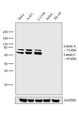
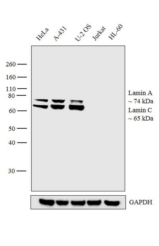
- Western blot analysis was performed on nuclear enriched cell extracts (30 µg lysate) of HeLa (Lane 1), A-431 (Lane 2), U-2 OS (Lane 3), Jurkat (Lane 4) and HL-60 (Lane 5). The blot was probed with Anti- Lamin A/C Antibody (Product # MA1-5820, 1:500 dilution) and detected by chemiluminescence using Goat anti-Mouse IgG (H+L) Superclonal™ Secondary Antibody, HRP conjugate (Product # A28177, 0.25 µg/mL, 1:4000 dilution). 74 and 65 kDa band corresponding to Lamin A and C respectively were observed across all the cell lines tested except Jurkat and HL-60.
Supportive validation
- Submitted by
- Invitrogen Antibodies (provider)
- Main image

- Experimental details
- Immunofluorescent analysis of Lamin A/C using monoclonal antibody (Product # MA1-5820).

Supportive validation
- Submitted by
- Invitrogen Antibodies (provider)
- Main image

- Experimental details
- Chromatin Immunoprecipitation-Western Blot (ChIP-WB) of endogenous Lamin A/C proteins using Anti-Lamin A/C Antibody: ChIP was performed using Lamin A/C Mouse Monoclonal Antibody (Product # MA1-5820, 10 µg) (lane 2) on sheared chromatin from 4 million formaldehyde fixed HeLa cells. Normal Rabbit IgG (lane 1) was used as negative IP control. Western blot analysis of immunoprecipitated proteins was performed using Lamin A Mouse Monoclonal Antibody (Product # MA1-06101, 1 µg/mL) and Lamin C Rabbit Polyclonal Antibody (Product # PA1-5827, 1:1000 dilution).

Supportive validation
- Submitted by
- Invitrogen Antibodies (provider)
- Main image

- Experimental details
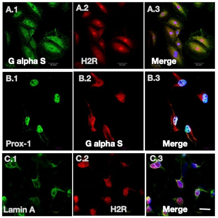
- Fig. 2. Colocalization of H2R with signaling partner G alpha S and nuclear envelope marker Lamin A in the nucleus. (A.1-3) Confocal images of LECs, labeled for G alpha S (green channel, A.1), H2R (red channel, A.2) and overlay merge of G alpha S, H2R and nuclear marker (DAPI) (A.3), showing the colocalization of H2R with signaling partner G alpha S in the nucleus. (B.1-3) LECs labeled with lymphatic specific marker Prox-1 (B.1), G alpha S (B.2) and overlay (B.3), showing expression of G alpha S in LECs. (C.1-3) Confocal images of nuclear envelope marker Lamin A (C.1) and H2R (C.2), demonstrating H2R localization in the nucleus in the overlay image (C.3). Scale bar: 20 um.