 Explore
 Validate
 Learn
LearnABIN1580428
antibody from antibodies-online
Targeting: LMNA
CMD1A, HGPS, LGMD1B, LMN1, LMNL1, MADA, PRO1
 Western blot
 Immunohistochemistry
Antibody data
- Antibody Data
- Antigen structure
- References [2]
- Comments [0]
- Validations
- Western blot [1]
- Immunocytochemistry [1]
Submit
Validation data
Reference
Comment
Report error
- Product number
- ABIN1580428 - Provider product page

- Provider
- antibodies-online
- Product name
- anti-Lamin A/C (LMNA) antibody
- Antibody type
- Polyclonal
- Antigen
- Other
- Description
- IgY preparation
- Reactivity
- Human, Mouse, Rat, Bovine, Porcine
- Host
- Chicken/Avian
- Isotype
- IgY
- Vial size
- 100 μL
- Concentration
- 20 mg/mL
- Storage
- Store at 4°C short term or -20°C long term.
- Handling
- Avoid repeated freezing and thawing.
Submitted references Lamin A/C, laminopathies and premature ageing.
cDNA sequencing of nuclear lamins A and C reveals primary and secondary structural homology to intermediate filament proteins.
Liu B, Zhou Z
Histology and histopathology 2008 Jun;23(6):747-63
Histology and histopathology 2008 Jun;23(6):747-63
cDNA sequencing of nuclear lamins A and C reveals primary and secondary structural homology to intermediate filament proteins.
Fisher DZ, Chaudhary N, Blobel G
Proceedings of the National Academy of Sciences of the United States of America 1986 Sep;83(17):6450-4
Proceedings of the National Academy of Sciences of the United States of America 1986 Sep;83(17):6450-4
No comments: Submit comment
Supportive validation
- Submitted by
- antibodies-online (provider)
- Main image
- Experimental details
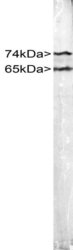
- Stripe blot of crude HeLa cell extract stained with CPCA-LaminAC. Note two strong clean bands at 74 kDa and 65 kDa, corresponding to Lamins A and C.

Supportive validation
- Submitted by
- antibodies-online (provider)
- Main image

- Experimental details
- HeLa cells staining with CPCA-LaminAC (red), and counterstained with monoclonal antibody to Lysosomal Associated Membrane Protein 1 (Lamp1), MCA- 6E2 (green) and DNA (blue). The CPCA-LaminAC antibody reveals strong nuclear lamina staining, while MCA- 6E2 antibody reveals strong cytoplasmic punctate staining of lysosomes and early endosomes. Since both DNA (blue) and Lamin A/C (red) are associated with the nuclear compartment, this region appears crimson in this image.
