 Explore
 Validate
 Learn
Learn Western blot
Antibody data
- Antibody Data
- Antigen structure
- References [1]
- Comments [0]
- Validations
- Western blot [5]
- Immunocytochemistry [1]
- Immunoprecipitation [1]
- Immunohistochemistry [6]
Submit
Validation data
Reference
Comment
Report error
- Product number
- GTX101126 - Provider product page

- Provider
- GeneTex
- Proper citation
- GeneTex Cat#GTX101126, RRID:AB_10617300
- Product name
- Lamin A + C antibody
- Antibody type
- Polyclonal
- Reactivity
- Human, Mouse
- Host
- Rabbit
Submitted references The histone demethylase jumonji coordinates cellular senescence including secretion of neural stem cell-attracting cytokines.
Perrigue PM, Silva ME, Warden CD, Feng NL, Reid MA, Mota DJ, Joseph LP, Tian YI, Glackin CA, Gutova M, Najbauer J, Aboody KS, Barish ME
Molecular cancer research : MCR 2015 Apr;13(4):636-50
Molecular cancer research : MCR 2015 Apr;13(4):636-50
No comments: Submit comment
Supportive validation
- Submitted by
- GeneTex (provider)
- Main image
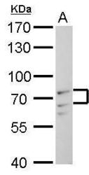
- Experimental details
- Lamin A + C antibody detects Lamin A + C protein by Western blot analysis.A. 30 £gg C2C12 whole cell lysate/extract7.5 % SDS-PAGELamin A + C antibody (GTX101126) dilution: 1:1000
- Submitted by
- GeneTex (provider)
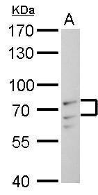
- Main image
- Experimental details
- Lamin A + C antibody detects Lamin A + C protein by Western blot analysis.A. 30 £gg HeLa whole cell lysate/extractB. 30 £gg HeLa nuclear lysate/extract7.5 % SDS-PAGELamin A + C antibody (GTX101126) dilution: 1:5000

- Submitted by
- GeneTex (provider)
- Main image
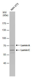
- Experimental details
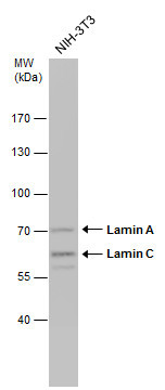
- Whole cell extract (30 £gg) was separated by 7.5% SDS-PAGE, and the membrane was blotted with Lamin A + C antibody (GTX101126) diluted at 1:1000.
- Submitted by
- GeneTex (provider)
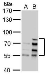
- Main image
- Experimental details
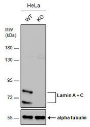
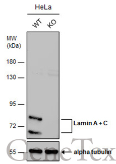
- Wild-type (WT) and Lamin A + C knockout (KO) HeLa cell extracts (30 ?g) were separated by 7.5% SDS-PAGE, and the membrane was blotted with Lamin A + C antibody (GTX101126) diluted at 1:5000. The HRP-conjugated anti-rabbit IgG antibody (GTX213110-01) was used to detect the primary antibody, and the signal was developed with Trident ECL plus-Enhanced.
- Submitted by
- GeneTex (provider)
- Main image
- Experimental details
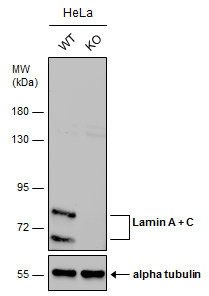
- Wild-type (WT) and Lamin A + C knockout (KO) HeLa cell extracts (30 ?g) were separated by 7.5% SDS-PAGE, and the membrane was blotted with Lamin A + C antibody (GTX101126) diluted at 1:5000. The HRP-conjugated anti-rabbit IgG antibody (GTX213110-01) was used to detect the primary antibody, and the signal was developed with Trident ECL plus-Enhanced.
Supportive validation
- Submitted by
- GeneTex (provider)
- Main image
- Experimental details
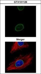
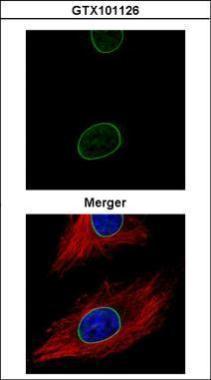
- Confocal immunofluorescence analysis (Olympus FV10i) of methanol-fixed HeLa, using Lamin A + C (GTX101126) antibody (Green) at 1:500 dilution. Alpha-tubulin filaments were labeled with GTX11304 (Red) at 1:500.
Supportive validation
- Submitted by
- GeneTex (provider)
- Main image
- Experimental details
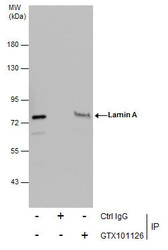
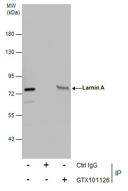
- Immunoprecipitation of Lamin A + C protein from HeLa whole cell extracts using 5 £gg of Lamin A + C antibody (GTX101126).Western blot analysis was performed using Lamin A + C antibody (GTX101126).EasyBlot anti-Rabbit IgG (GTX221666-01) was used as a secondary reagent.
Supportive validation
- Submitted by
- GeneTex (provider)
- Main image
- Experimental details
- Lamin A + C antibody detects Lamin A + C protein at membrane on U87 xenograft by immunohistochemical analysis. Sample: Paraffin-embedded U87 xenograft. Lamin A + C antibody (GTX101126) dilution: 1:500.
- Submitted by
- GeneTex (provider)
- Main image
- Experimental details
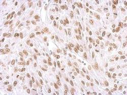
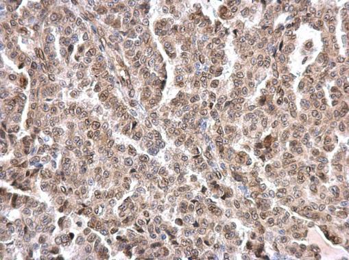
- Lamin A + C antibody detects Lamin A + C protein at nuclear envelope on human breast carcinoma by immunohistochemical analysis. Sample: Paraffin-embedded human breast carcinoma. Lamin A + C antibody (GTX101126) dilution: 1:500.

- Submitted by
- GeneTex (provider)
- Main image
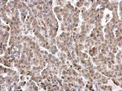
- Experimental details
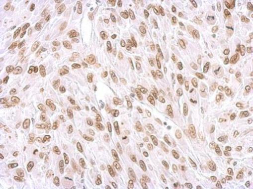
- Lamin A + C antibody detects Lamin A + C protein at nuclear envelope on human endometrial carcinoma by immunohistochemical analysis. Sample: Paraffin-embedded human endometrial carcinoma. Lamin A + C antibody (GTX101126) dilution: 1:500.
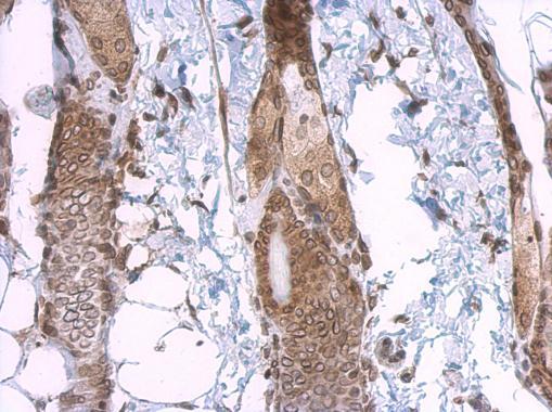
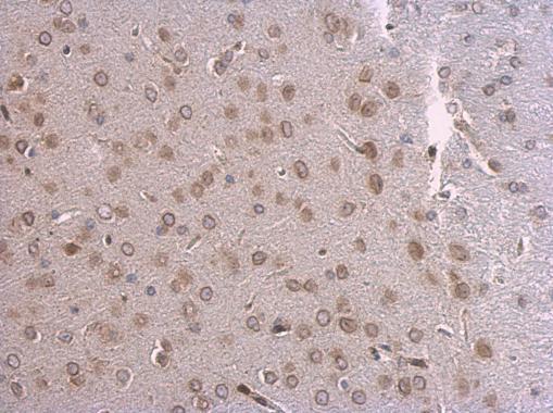
- Submitted by
- GeneTex (provider)
- Main image
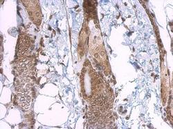
- Experimental details
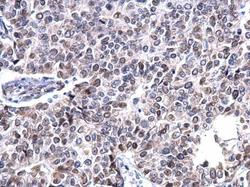
- Lamin A + C antibody detects Lamin A + C protein at nuclear envelope on mouse skin by immunohistochemical analysis. Sample: Paraffin-embedded mouse skin. Lamin A + C antibody (GTX101126) dilution: 1:500.
- Submitted by
- GeneTex (provider)
- Main image
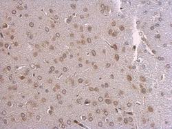
- Experimental details
- Lamin A + C antibody detects Lamin A + C protein at nuclear envelope on mouse fore brain by immunohistochemical analysis. Sample: Paraffin-embedded mouse fore brain. Lamin A + C antibody (GTX101126) dilution: 1:500.
- Submitted by
- GeneTex (provider)
- Main image
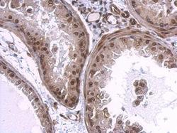
- Experimental details
- Lamin A + C antibody detects Lamin A + C protein at nuclear envelope on mouse prostate by immunohistochemical analysis. Sample: Paraffin-embedded mouse prostate. Lamin A + C antibody (GTX101126) dilution: 1:500.
