Explore
 Validate
 Learn
LearnSTJ11101676
antibody from St John's Laboratory
Targeting: LMNA
CMD1A, HGPS, LGMD1B, LMN1, LMNL1, MADA, PRO1
 Western blot
 Immunocytochemistry Immunoprecipitation Immunohistochemistry Chromatin Immunoprecipitation
Antibody data
- Antibody Data
- Antigen structure
- References [0]
- Comments [0]
- Validations
- Western blot [4]
- Immunohistochemistry [6]
- Chromatin Immunoprecipitation [1]
Submit
Validation data
Reference
Comment
Report error
- Product number
- STJ11101676 - Provider product page

- Provider
- St John's Laboratory
- Product name
- Anti-LMNA antibody [ARC5001] (STJ11101676)
- Antibody type
- Monoclonal
- Description
- Rabbit monoclonal antibody anti-Lamin A/C is suitable for use in Western Blot, Immunohistochemistry, Immunofluorescence and Immunoprecipitation.
- Reactivity
- Human, Mouse, Rat
- Host
- Rabbit
- Conjugate
- Unconjugated
- Antigen sequence
NA- Epitope
- NA
- Isotype
- IgG
- Antibody clone number
- NA
- Vial size
- NA
- Concentration
- NA
- Storage
- Store in a freezer at-20°C and avoid freeze-thaw cycles.
- Handling
- NA
No comments: Submit comment
Supportive validation
Supportive validation
Supportive validation
Supportive validation
- Submitted by
- St John's Laboratory (provider)
- Main image
- Experimental details
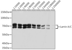
- Western blot analysis of extracts of HeLa cells, using Lamin A/C antibody (STJ11101676) at 1:10000-1:2560000 dilution. Secondary antibody: HRP Goat Anti-rabbit IgG (H+L) at 1:10000 dilution. Lysates/proteins: 25ug per lane. Blocking buffer: 3% nonfat dry milk in TBST. Detection: ECL Basic Kit. Exposure time: 1s.
- Sample type
- NA
- Validation comment
- NA
- Primary Ab dilution
- NA
- Other comments
- NA
- Secondary Ab
- NA
- Secondary Ab dilution
- NA
- Protocol
- NA

Supportive validation
- Submitted by
- St John's Laboratory (provider)
- Main image

- Experimental details
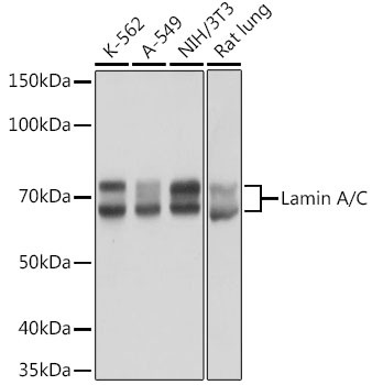
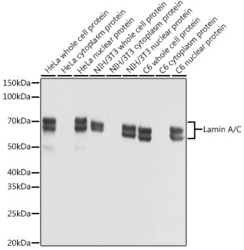
- Western blot analysis of extracts of various cell lines, using Lamin A/C antibody (STJ11101676) at 1:50000 dilution. Secondary antibody: HRP Goat Anti-rabbit IgG (H+L) at 1:10000 dilution. Lysates/proteins: 25ug per lane. Blocking buffer: 3% nonfat dry milk in TBST. Detection: ECL Basic Kit. Exposure time: 1s.
- Sample type
- NA
- Validation comment
- NA
- Primary Ab dilution
- NA
- Other comments
- NA
- Secondary Ab
- NA
- Secondary Ab dilution
- NA
- Protocol
- NA
Supportive validation
- Submitted by
- St John's Laboratory (provider)
- Main image

- Experimental details
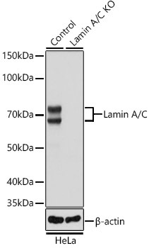
- Western blot analysis of extracts from normal (control) and Lamin A/C knockout (KO) HeLa cells, using Lamin A/C antibody (STJ11101676) at 1:50000 dilution. Secondary antibody: HRP Goat Anti-rabbit IgG (H+L) at 1:10000 dilution. Lysates/proteins: 25ug per lane. Blocking buffer: 3% nonfat dry milk in TBST. Detection: ECL Basic Kit. Exposure time: 1s.
- Sample type
- NA
- Validation comment
- NA
- Primary Ab dilution
- NA
- Other comments
- NA
- Secondary Ab
- NA
- Secondary Ab dilution
- NA
- Protocol
- NA
Supportive validation
- Submitted by
- St John's Laboratory (provider)
- Main image
- Experimental details
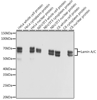
- Western blot analysis of extracts of various cell lines, using Lamin A/C antibody (STJ11101676) at 1:10000 dilution. Secondary antibody: HRP Goat Anti-rabbit IgG (H+L) at 1:10000 dilution. Lysates/proteins: 25ug per lane. Blocking buffer: 3% nonfat dry milk in TBST. Detection: ECL Basic Kit. Exposure time: 1s.
- Sample type
- NA
- Validation comment
- NA
- Primary Ab dilution
- NA
- Other comments
- NA
- Secondary Ab
- NA
- Secondary Ab dilution
- NA
- Protocol
- NA
Supportive validation
Supportive validation
Supportive validation
Supportive validation
Supportive validation
Supportive validation
- Submitted by
- St John's Laboratory (provider)
- Main image
- Experimental details
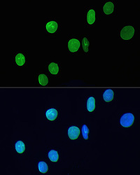
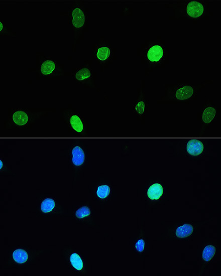
- Immunofluorescence analysis of H9C2 cells using Lamin A/C antibody (STJ11101676) at dilution of 1:200. Blue: DAPI for nuclear staining.
- Sample type
- NA
- Validation comment
- NA
- Primary Ab dilution
- NA
- Other comments
- NA
- Secondary Ab
- NA
- Secondary Ab dilution
- NA
- Protocol
- NA

Supportive validation
- Submitted by
- St John's Laboratory (provider)
- Main image
- Experimental details
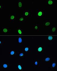
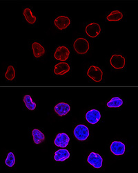
- Immunofluorescence analysis of L929 cells using Lamin A/C antibody (STJ11101676) at dilution of 1:200. Blue: DAPI for nuclear staining.
- Sample type
- NA
- Validation comment
- NA
- Primary Ab dilution
- NA
- Other comments
- NA
- Secondary Ab
- NA
- Secondary Ab dilution
- NA
- Protocol
- NA
Supportive validation
- Submitted by
- St John's Laboratory (provider)
- Main image
- Experimental details
- Confocal immunofluorescence analysis of HeLa cells using Lamin A/C antibody (STJ11101676) at dilution of 1:100. Blue: DAPI for nuclear staining.
- Sample type
- NA
- Validation comment
- NA
- Primary Ab dilution
- NA
- Other comments
- NA
- Secondary Ab
- NA
- Secondary Ab dilution
- NA
- Protocol
- NA

Supportive validation
- Submitted by
- St John's Laboratory (provider)
- Main image
- Experimental details
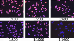
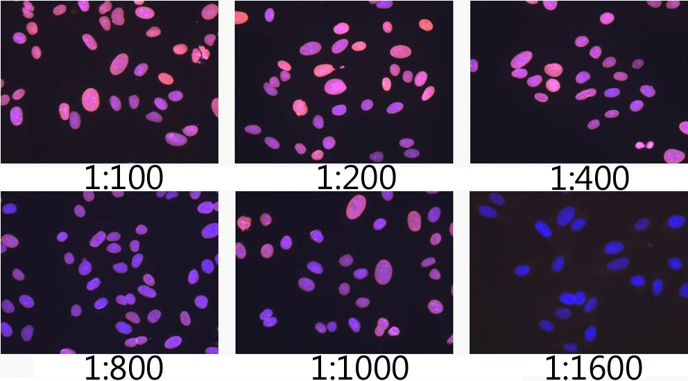
- Immunofluorescence analysis of U2OS cells using Lamin A/C antibody (STJ11101676) at dilution of 1:100- 1:1600. Blue: DAPI for nuclear staining.
- Sample type
- NA
- Validation comment
- NA
- Primary Ab dilution
- NA
- Other comments
- NA
- Secondary Ab
- NA
- Secondary Ab dilution
- NA
- Protocol
- NA
Supportive validation
- Submitted by
- St John's Laboratory (provider)
- Main image
- Experimental details
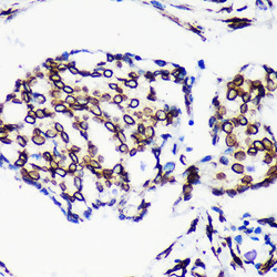
- Immunohistochemistry of paraffin-embedded Human breast cancer using Lamin A/C antibody (STJ11101676) at dilution of 1:200 (40x lens). Perform microwave antigen retrieval with 10 mM PBS buffer pH 7. 2 before commencing with immunohistochemistry staining protocol.
- Sample type
- NA
- Validation comment
- NA
- Primary Ab dilution
- NA
- Other comments
- NA
- Secondary Ab
- NA
- Secondary Ab dilution
- NA
- Protocol
- NA

Supportive validation
- Submitted by
- St John's Laboratory (provider)
- Main image
- Experimental details
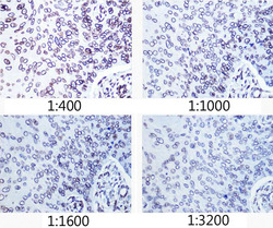
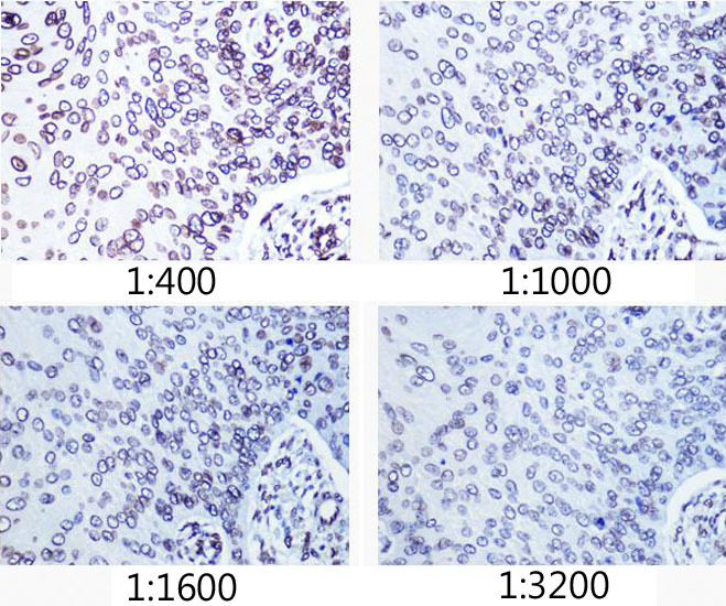
- Immunohistochemistry of paraffin-embedded human esophageal cancer using Lamin A/C antibody (STJ11101676) at dilution of 1:400- 1:3200 (40x lens). Perform microwave antigen retrieval with 10 mM PBS buffer pH 7. 2 before commencing with immunohistochemistry staining protocol.
- Sample type
- NA
- Validation comment
- NA
- Primary Ab dilution
- NA
- Other comments
- NA
- Secondary Ab
- NA
- Secondary Ab dilution
- NA
- Protocol
- NA
Supportive validation
- Submitted by
- St John's Laboratory (provider)
- Main image

- Experimental details
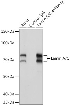
- Immunoprecipitation analysis of 200ug extracts of K-562 cells using 3ug Lamin A/C antibody (STJ11101676). Western blot was performed from the immunoprecipitate using Lamin A/C antibody (STJ11101676) at a dilition of 1:20000.
- Sample type
- NA
- Validation comment
- NA
- Primary Ab dilution
- NA
- Other comments
- NA
- Secondary Ab
- NA
- Secondary Ab dilution
- NA
- Protocol
- NA