 Explore
 Validate
 Learn
Learn Western blot
Antibody data
- Antibody Data
- Antigen structure
- References [0]
- Comments [0]
- Validations
- Western blot [1]
- Immunocytochemistry [2]
- Immunohistochemistry [1]
Submit
Validation data
Reference
Comment
Report error
- Product number
- APC-035-GP-25UL - Provider product page

- Provider
- Invitrogen Antibodies
- Product name
- Kir4.1 (KCNJ10) Polyclonal Antibody
- Antibody type
- Polyclonal
- Antigen
- Other
- Reactivity
- Human, Mouse, Rat
- Host
- Guinea Pig
- Isotype
- IgG
- Vial size
- 25 µL
- Concentration
- 0.8 mg/mL
- Storage
- -20° C, Avoid Freeze/Thaw Cycles
No comments: Submit comment
Supportive validation
- Submitted by
- Invitrogen Antibodies (provider)
- Main image
- Experimental details
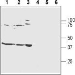
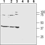
- Western blot analysisof rat brain membranes (lanes 1 and 4), mouse brain membranes (lanes 2 and 5) and human SH-SY5Y neuroblastoma cell lysates (lanes 3 and 6): - 1-3.Guinea pig Anti-Kir4.1 (KCNJ10) Antibody (#APC-035-GP), (1:800).4-6. Guinea pig Anti-Kir4.1 (KCNJ10) Antibody , preincubated with Kir4.1/KCNJ10 Blocking Peptide (#BLP-PC035).
Supportive validation
- Submitted by
- Invitrogen Antibodies (provider)
- Main image

- Experimental details
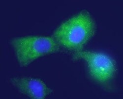
- Expression of Kir4.1 in human U-87 MG glioblastoma cell line - Immunocytochemical staining of fixed and permeabilized U-87 MG cells using Guinea pig Anti-Kir4.1 (KCNJ10) Antibody (#APC-035-GP), (1:100) followed by goat Anti-guinea pig-AlexaFluor-488 secondary Antibody (green). Cell nuclei were visualized using Hoechst 33342 (blue).

- Submitted by
- Invitrogen Antibodies (provider)
- Main image
- Experimental details
- Expression of Kir4.1 in human U-87 MG glioblastoma cell line - Immunocytochemical staining of fixed and permeabilized U-87 MG cells using Guinea pig Anti-Kir4.1 (KCNJ10) Antibody (#APC-035-GP), (1:100) followed by goat Anti-guinea pig-AlexaFluor-488 secondary Antibody (green). Cell nuclei were visualized using Hoechst 33342 (blue).

Supportive validation
- Submitted by
- Invitrogen Antibodies (provider)
- Main image
- Experimental details
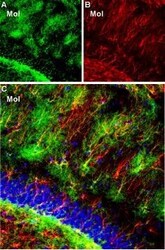
- Expression of Kir4.1 in rat hippocampal dentate gyrus - Immunohistochemical staining of rat frozen brain sections using Guinea pig Anti-Kir4.1 (KCNJ10) Antibody (#APC-035-GP), (1:300). A.Kir4.1 staining (green) reveals Kir4.1 clustersin the molecular layer (Mol). B. The same section was stained with mouse Anti-glial fibrillary acidic protein (GFAP), (red) showing astrocyte profiles. C. Merge of Kir4.1 and GFAP images reveals colocalization on some branches of astrocytic fibers. In addition, Kir4.1 is expressed in the fine ramification of astrocytic fibers, thus creating a “halo-like” cloud around the GFAP astrocytic outline. DAPI (blue) counterstain reveals the outline of the dentate granule layer.
