Explore
 Validate
 Learn
Learn Western blot
 Other assay
Antibody data
- Antibody Data
- Antigen structure
- References [1]
- Comments [0]
- Validations
- Other assay [2]
Submit
Validation data
Reference
Comment
Report error
- Product number
- PA5-28284 - Provider product page

- Provider
- Invitrogen Antibodies
- Product name
- KCNMB1 Polyclonal Antibody
- Antibody type
- Polyclonal
- Antigen
- Recombinant full-length protein
- Description
- Recommended positive controls: 293T, A431, H1299, HeLa, HepG2, Molt-4, Raji, mouse heart. Predicted reactivity: Mouse (80%), Rat (81%), Dog (84%), Rabbit (85%), Rhesus Monkey (94%), Bovine (84%). Store product as a concentrated solution. Centrifuge briefly prior to opening the vial.
- Reactivity
- Human, Mouse
- Host
- Rabbit
- Isotype
- IgG
- Vial size
- 100 μL
- Concentration
- 1 mg/mL
- Storage
- Store at 4°C short term. For long term storage, store at -20°C, avoiding freeze/thaw cycles.
Submitted references Western blot analysis of BK channel β1-subunit expression should be interpreted cautiously when using commercially available antibodies.
Bhattarai Y, Fernandes R, Kadrofske MM, Lockwood LR, Galligan JJ, Xu H
Physiological reports 2014 Oct 1;2(10)
Physiological reports 2014 Oct 1;2(10)
No comments: Submit comment
Supportive validation
- Submitted by
- Invitrogen Antibodies (provider)
- Main image

- Experimental details
- NULL
- Submitted by
- Invitrogen Antibodies (provider)
- Main image

- Experimental details
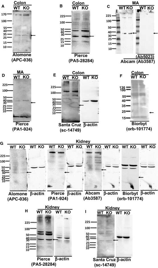
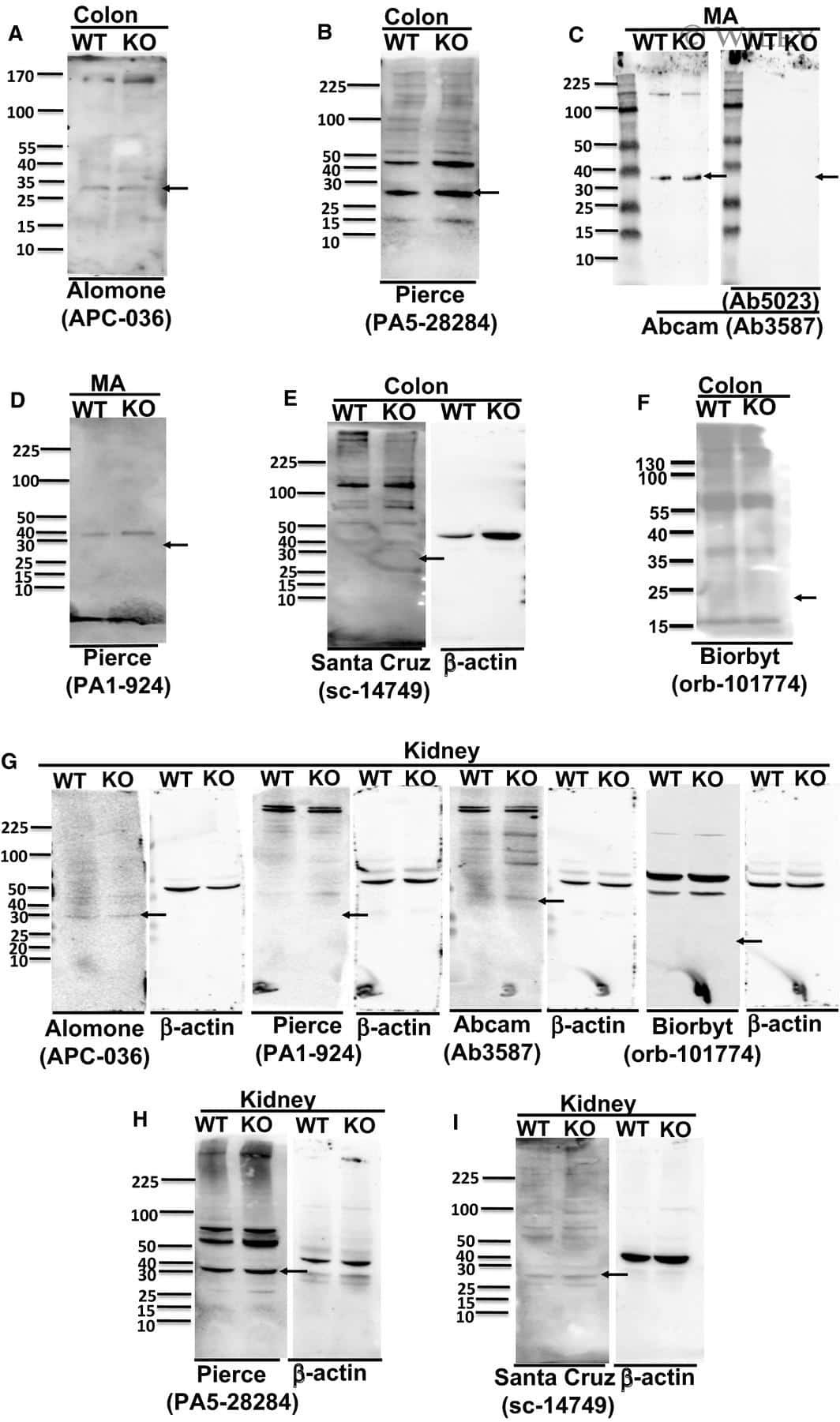
- Figure 1. Representative western blot obtained using anti-BK beta 1-subunit antibodies in MA, colonic, or kidney tissues from WT and BK beta 1-KO mice, (A) Alomone Labs (APC-036), (B) Pierce (PA5-28284), and (C) Abcam (Ab3587) antibodies detected a protein band at ~28 kDa or ~38 kDa in colons or MA from both mice. The bands in Abcam sets were diminished after preincubation of the primary antibody with the competing peptide. (D) Pierce (PA1-924), (E) Santa Cruz (sc-14749), and (F) Biorbyt (orb-101774) antibodies did not detect any band at ~28 kDa or ~21 kDa in MA or colons from WT mice. (G) Alomone Labs (APC-036), Pierce (PA1-924), Abcam (Ab3587), and Biorbyt (Orb-101774) antibodies did not detect protein band at ~28 kDa, ~38 kDa, or ~21 kDa in kidneys from WT mice. (H) Pierce (PA5-28284) and (I) Santa Cruz (sc-14749) antibodies detected the protein band at ~28 kDa in kidneys from both mice. beta -actin was reblotted on each membrane after anti-BK beta 1-subunit antibody was stripped. All representative blot images from kidney are in the tissue from same WT or BK beta 1-KO mouse, and blotted with primary anti-BK beta 1-subunit antibody at 1:200. Arrows indicate the manufacturer's recommended molecular weight of BK beta 1-subunit protein.