Explore
 Validate
 Learn
Learn Western blot
 Immunocytochemistry
 Immunoprecipitation
Antibody data
- Antibody Data
- Antigen structure
- References [2]
- Comments [0]
- Validations
- Immunocytochemistry [2]
- Other assay [2]
Submit
Validation data
Reference
Comment
Report error
- Product number
- PA1-969 - Provider product page

- Provider
- Invitrogen Antibodies
- Product name
- PSMC2 Polyclonal Antibody
- Antibody type
- Polyclonal
- Antigen
- Recombinant full-length protein
- Description
- PA1-969 detects proteasome 19S subunit S7 from human cells. PA1-969 has been successfully used in Western blot and immunoprecipitation procedures. By Western blot, this antibody detects a 47 kDa protein representing proteasome 19S subunit S7 from HeLa cell lysate. PA1-969 antigen is recombinant human proteasome 19S subunit S7.
- Reactivity
- Human
- Host
- Rabbit
- Isotype
- IgG
- Vial size
- 100 μL
- Concentration
- Conc. Not Determined
- Storage
- -20°C, Avoid Freeze/Thaw Cycles
Submitted references PSMC2 is overexpressed in glioma and promotes proliferation and anti-apoptosis of glioma cells.
The cyclophilin-like domain of Ran-binding protein-2 modulates selectively the activity of the ubiquitin-proteasome system and protein biogenesis.
Zheng X, Wang Y, Wang D, Wan J, Qin X, Mu Z, Hu N
World journal of surgical oncology 2022 Mar 14;20(1):84
World journal of surgical oncology 2022 Mar 14;20(1):84
The cyclophilin-like domain of Ran-binding protein-2 modulates selectively the activity of the ubiquitin-proteasome system and protein biogenesis.
Yi H, Friedman JL, Ferreira PA
The Journal of biological chemistry 2007 Nov 30;282(48):34770-8
The Journal of biological chemistry 2007 Nov 30;282(48):34770-8
No comments: Submit comment
Supportive validation
- Submitted by
- Invitrogen Antibodies (provider)
- Main image
- Experimental details
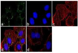
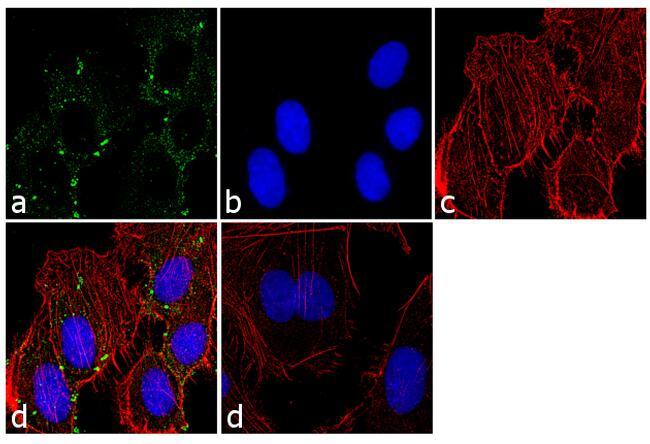
- Immunofluorescence analysis of PSMC2 was performed using 70% confluent log phase SK-OV-3 cells. The cells were fixed with 4% paraformaldehyde for 10 minutes, permeabilized with 0.1% Triton™ X-100 for 10 minutes, and blocked with 1% BSA for 1 hour at room temperature. The cells were labeled with PSMC2 Rabbit Polyclonal Antibody (Product # PA1-969) at 1:250 dilution in0.1% BSA and incubated for 3 hours at room temperature and then labeled with Goat anti-Rabbit IgG (H+L) Superclonal™ Secondary Antibody, Alexa Fluor® 488 conjugate (Product # A27034) at a dilution of 1:2000 for 45 minutes at room temperature (Panel a: green). Nuclei (Panel b: blue) were stained with SlowFade® Gold Antifade Mountant with DAPI (Product # S36938). F-actin (Panel c: red) was stained with Rhodamine Phalloidin (Product # R415, 1:300). Panel d represents the merged image showing cytoplasmic localization. Panel e shows the control without primary antibody. The images were captured at 60X magnification.

- Submitted by
- Invitrogen Antibodies (provider)
- Main image
- Experimental details
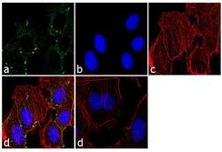
- Immunofluorescence analysis of PSMC2 was performed using 70% confluent log phase SK-OV-3 cells. The cells were fixed with 4% paraformaldehyde for 10 minutes, permeabilized with 0.1% Triton™ X-100 for 10 minutes, and blocked with 1% BSA for 1 hour at room temperature. The cells were labeled with PSMC2 Rabbit Polyclonal Antibody (Product # PA1-969) at 1:250 dilution in0.1% BSA and incubated for 3 hours at room temperature and then labeled with Goat anti-Rabbit IgG (Heavy Chain) Superclonal™ Secondary Antibody, Alexa Fluor® 488 conjugate (Product # A27034) at a dilution of 1:2000 for 45 minutes at room temperature (Panel a: green). Nuclei (Panel b: blue) were stained with SlowFade® Gold Antifade Mountant with DAPI (Product # S36938). F-actin (Panel c: red) was stained with Rhodamine Phalloidin (Product # R415, 1:300). Panel d represents the merged image showing cytoplasmic localization. Panel e shows the control without primary antibody. The images were captured at 60X magnification.
Supportive validation
- Submitted by
- Invitrogen Antibodies (provider)
- Main image
- Experimental details
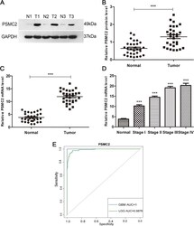
- Expression of PSMC2 in glioma. A The protein levels of PSMC2 in three pairs of glioma tissues (T) and adjacent non-tumor tissues (N) measured by Western blot. B PSMC2 protein levels were quantified in glioma ( n = 30) and normal control ( n = 30) tissues. C mRNA levels of PSMC2 in 30 glioma tissues and 30 normal control tissues. D mRNA levels of PSMC2 in gliomas of different malignant grades, in which grade I, n = 5; grade II, n = 8; grade III, n = 7; and grade IV, n = 10. T, glioma tissue; N, normal control tissue (*** p < 0.001). E ROC curves of differential expression of PSMC2 gene in cancer tissues (GBM, LGG) and normal tissues in the joint analysis of TCGA and GTEX databases
- Submitted by
- Invitrogen Antibodies (provider)
- Main image
- Experimental details
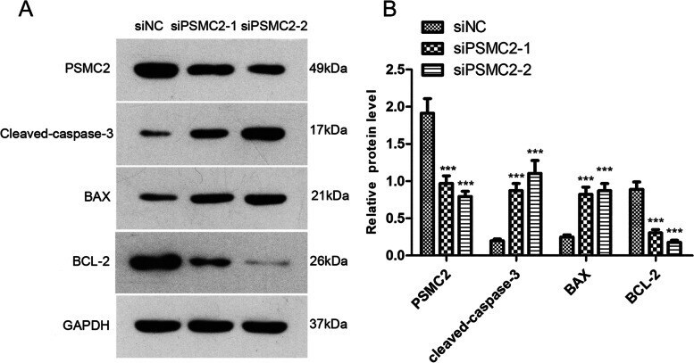
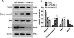
- Effect of PSMC2 on apoptosis-related proteins in glioma cells. A Western blot of PSMC2 and apoptosis pathway-related proteins after transfection of PSMC2 siRNA into U343MG cells. B Statistical results of PSMC2 and apoptosis pathway-related proteins after transfection of PSMC2 siRNA into U343MG cells