 Explore
 Validate
 Learn
Learn Flow cytometry
Antibody data
- Antibody Data
- Antigen structure
- References [3]
- Comments [0]
- Validations
- Flow cytometry [1]
Submit
Validation data
Reference
Comment
Report error
- Product number
- 12-5846-80 - Provider product page

- Provider
- Invitrogen Antibodies
- Product name
- Anti-TdT Monoclonal Antibody (19-3), PE, eBioscience™
- Antibody type
- Monoclonal
- Antigen
- Other
- Description
- Description: This 19-3 monoclonal antibody recognizes human and mouse terminal-deoxynucleotidyl transferase (TdT), a 60 kDa polymerase responsible for the template-independent addition of N-nucleotides at gene segment junctions of developing lymphocytes. Expression is found in the nucleus of immature lymphocytes but not in mature lymphocytes or non-lymphoid cells. Tdt plays a critical role during TCR and Ig gene rearrangement. Regulation of expression in the mouse thymus correlates with T cell selection; decreased or absent after positive selection. Additionally Tdt has been shown to be present in lymphoma or lymphoblastic leukemia cells. Applications Reported: This 19-3 antibody has been reported for use in intracellular staining followed by flow cytometric analysis. Applications Tested: This 19-3 antibody has been tested by intracellular staining using the Foxp3/Transcription Factor Staining Buffer Set (cat. 00-5521) followed by flow cytometric analysis of mouse thymocytes. This can be used at less than or equal to 0.5 µg per test. A test is defined as the amount (µg) of antibody that will stain a cell sample in a final volume of 100 µL. Cell number should be determined empirically but can range from 10^5 to 10^8 cells/test. It is recommended that the antibody be carefully titrated for optimal performance in the assay of interest. Excitation: 488-561 nm; Emission: 578 nm; Laser: Blue Laser, Green Laser, Yellow-Green Laser. Filtration: 0.2 µm post-manufacturing filtered.
- Reactivity
- Human, Mouse
- Host
- Mouse
- Conjugate
- Yellow dye
- Isotype
- IgG
- Antibody clone number
- 19-3
- Vial size
- 25 µg
- Concentration
- 0.2 mg/mL
- Storage
- 4° C, store in dark, DO NOT FREEZE!
Submitted references Regulation of N-region diversity in antigen receptors through thymocyte differentiation and thymus ontogeny.
Detection of terminal deoxynucleotidyl transferase (TdT) by flow cytometry in leukemic disorders.
Immunological detection of a conserved structure for terminal deoxynucleotidyltransferase.
Bogue M, Gilfillan S, Benoist C, Mathis D
Proceedings of the National Academy of Sciences of the United States of America 1992 Nov 15;89(22):11011-5
Proceedings of the National Academy of Sciences of the United States of America 1992 Nov 15;89(22):11011-5
Detection of terminal deoxynucleotidyl transferase (TdT) by flow cytometry in leukemic disorders.
Bardales RH, Carrato A, Fleischer M, Schwartz MK, Koziner B
The journal of histochemistry and cytochemistry : official journal of the Histochemistry Society 1989 Apr;37(4):509-13
The journal of histochemistry and cytochemistry : official journal of the Histochemistry Society 1989 Apr;37(4):509-13
Immunological detection of a conserved structure for terminal deoxynucleotidyltransferase.
Bollum FJ, Chang LM
The Journal of biological chemistry 1981 Aug 25;256(16):8767-70
The Journal of biological chemistry 1981 Aug 25;256(16):8767-70
No comments: Submit comment
Supportive validation
- Submitted by
- Invitrogen Antibodies (provider)
- Main image
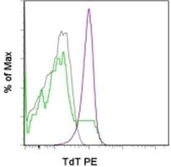
- Experimental details
- Staining of C57BL/6 thymocytes with Anti-Mouse CD4 FITC (Product # 11-0041-82) and Anti-Mouse CD8a PerCP-Cy5-5 (Product # 45-0081-82) followed by fixation and permeabilization with the Foxp3 Staining Buffers (Product # 00-5523-00) and subsequent staining with 0.25 µg of Anti-Mouse TdT PE. CD4 single positives are shown in gray, CD8 single positives in green and double positives in purple.
- Conjugate
- Yellow dye
