Explore
 Validate
 Learn
Learn Western blot
Antibody data
- Antibody Data
- Antigen structure
- References [4]
- Comments [0]
- Validations
- Western blot [1]
- Immunohistochemistry [1]
Submit
Validation data
Reference
Comment
Report error
- Product number
- 14-9739-80 - Provider product page

- Provider
- Invitrogen Antibodies
- Product name
- TdT Monoclonal Antibody (7UNA8U6), eBioscience™
- Antibody type
- Monoclonal
- Antigen
- Other
- Description
- Description: The monoclonal antibody 7UNA8U6 reacts with human, mouse, and rat TdT. TdT, or terminal-deoxynucleotidyl transferase, is a 60 kDa polymerase responsible for the template-independent addition of N-nucleotides at gene segment junctions of developing lymphocytes. Expression is found in the nucleus of immature lymphocytes but not in mature lymphocytes or non-lymphoid cells. TdT plays a critical role during TCR and Ig gene rearrangement and functions to increase the diversity of the lymphocyte repertoire through the addition of N-nucleotides onto the ends of newly formed P-nucleotides. After positive selection, expression of TdT becomes decreased or absent. Expression of TdT has been shown to be present in lymphoma or lymphoblastic leukemia cells. Applications Reported: This 7UNA8U6 antibody has been reported for use in western blotting and immunohistochemical staining of formalin-fixed paraffin embedded tissue sections. Applications Tested: This 7UNA8U6 antibody has been tested by immunohistochemistry on formalin-fixed paraffin embedded human and mouse tissue using low pH antigen retrieval and can be used at less than or equal to 10 µg/mL. This 7UNA8U6 antibody has also been tested by western blot analysis of reduced cell lysate prepared from mouse and rat cells and can be used at less than or equal to 5 µg/mL. It is recommended that the antibody be carefully titrated for optimal performance in the assay of interest. Purity: Greater than 90%, as determined by SDS-PAGE. Aggregation: Less than 10%, as determined by HPLC. Filtration: 0.2 µm post-manufacturing filtered.
- Reactivity
- Human, Mouse, Rat
- Host
- Mouse
- Isotype
- IgG
- Antibody clone number
- 7UNA8U6
- Vial size
- 25 µg
- Concentration
- 0.5 mg/mL
- Storage
- 4° C
Submitted references Lack of N regions in antigen receptor variable region genes of TdT-deficient lymphocytes.
Regulation of N-region diversity in antigen receptors through thymocyte differentiation and thymus ontogeny.
Clinical utility of leukemia cell terminal transferase measurements.
Immunological detection of a conserved structure for terminal deoxynucleotidyltransferase.
Komori T, Okada A, Stewart V, Alt FW
Science (New York, N.Y.) 1993 Aug 27;261(5125):1171-5
Science (New York, N.Y.) 1993 Aug 27;261(5125):1171-5
Regulation of N-region diversity in antigen receptors through thymocyte differentiation and thymus ontogeny.
Bogue M, Gilfillan S, Benoist C, Mathis D
Proceedings of the National Academy of Sciences of the United States of America 1992 Nov 15;89(22):11011-5
Proceedings of the National Academy of Sciences of the United States of America 1992 Nov 15;89(22):11011-5
Clinical utility of leukemia cell terminal transferase measurements.
McCaffrey R, Lillquist A, Sallan S, Cohen E, Osband M
Cancer research 1981 Nov;41(11 Pt 2):4814-20
Cancer research 1981 Nov;41(11 Pt 2):4814-20
Immunological detection of a conserved structure for terminal deoxynucleotidyltransferase.
Bollum FJ, Chang LM
The Journal of biological chemistry 1981 Aug 25;256(16):8767-70
The Journal of biological chemistry 1981 Aug 25;256(16):8767-70
No comments: Submit comment
Supportive validation
- Submitted by
- Invitrogen Antibodies (provider)
- Main image
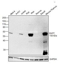
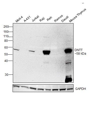
- Experimental details
- Western blot was performed using Anti-TdT Monoclonal Antibody (7UNA8U6), eBioscience™ (Product # 14-9739-80, 14-9739-82) and a 58 kDa band corresponding to DNA nucleotidylexotransferase was observed across positive cell lines (Molt-4, Jurkat and Reh) and tissue (Mouse thymus); and not across negative models (A-431, Raji, Ramos and Daudi). Whole cell extracts (30 µg lysate) of Molt-4 (Lane 1), A-431 (Lane 2), Jurkat (Lane 3), Raji (Lane 4), Reh (Lane 5), Ramos (Lane 6), Daudi (Lane 7) and tissue extract of Mouse Thymus (Lane 8) were electrophoresed using NuPAGE™ 10% Bis-Tris Protein Gel (Product # NP0301BOX). Resolved proteins were then transferred onto a Nitrocellulose membrane (Product # IB23001) by iBlot® 2 Dry Blotting System (Product # IB21001). The blot was probed with the primary antibody (5 µg/mL) and detected by chemiluminescence with Goat anti-Mouse IgG (H+L) Superclonal™ Recombinant Secondary Antibody, HRP (Product # A28177, 1:4000 dilution) using the iBright FL 1000 (Product # A32752). Chemiluminescent detection was performed using Novex® ECL Chemiluminescent Substrate Reagent Kit (Product # WP20005).
Supportive validation
- Submitted by
- Invitrogen Antibodies (provider)
- Main image

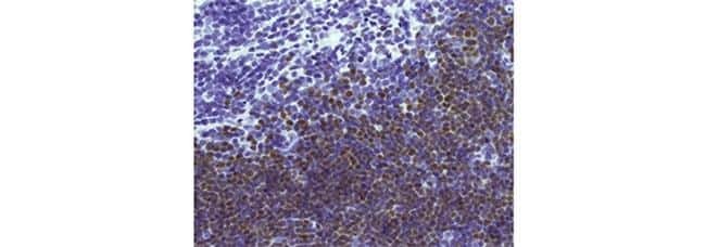
- Experimental details
- Immunohistochemistry of formalin-fixed paraffin embedded human thymus using 10 µg/mL of Anti-TdT Purified followed by Anti-Mouse Biotin, Streptavidin-HRP, and DAB visualization.Nuclei are counterstained with hematoxylin.