Explore
 Validate
 Learn
Learn Western blot
Antibody data
- Antibody Data
- Antigen structure
- References [0]
- Comments [0]
- Validations
- Western blot [4]
Submit
Validation data
Reference
Comment
Report error
- Product number
- PA5-28880 - Provider product page

- Provider
- Invitrogen Antibodies
- Product name
- TdT Polyclonal Antibody
- Antibody type
- Polyclonal
- Antigen
- Recombinant protein fragment
- Description
- Recommended positive controls: Jurkat, Jurkat nuclear, human DNTT-transfected 293T. Predicted reactivity: Mouse (80%), Rat (82%), Rhesus Monkey (98%), Bovine (88%). Store product as a concentrated solution. Centrifuge briefly prior to opening the vial.
- Reactivity
- Human
- Host
- Rabbit
- Isotype
- IgG
- Vial size
- 100 µL
- Concentration
- 1 mg/mL
- Storage
- Store at 4°C short term. For long term storage, store at -20°C, avoiding freeze/thaw cycles.
No comments: Submit comment
Supportive validation
- Submitted by
- Invitrogen Antibodies (provider)
- Main image

- Experimental details
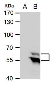
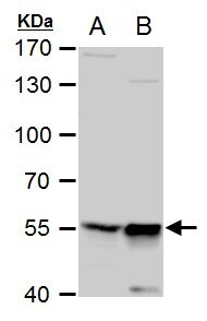
- Western blot analysis of TdT using A) 30 µg 293T whole cell extract and B) 30 µg whole cell extract of human DNTT-transfected 293T cells. Samples were loaded onto a 7.5% SDS-PAGE gel and probed with a TdT polyclonal antibody (Product # PA5-28880) at a dilution of 1:5000.
- Submitted by
- Invitrogen Antibodies (provider)
- Main image
- Experimental details
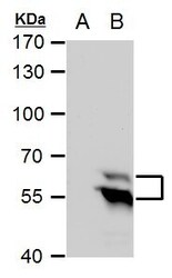
- TdT Polyclonal Antibody detects TdT protein by western blot analysis. A. 30 µg 293T whole cell extract. B. 30 µg whole cell extract of human DNTT-transfected 293T cells.7.5 % SDS-PAGE. TdT Polyclonal Antibody (Product # PA5-28880) dilution: 1:5,000.
- Submitted by
- Invitrogen Antibodies (provider)
- Main image

- Experimental details
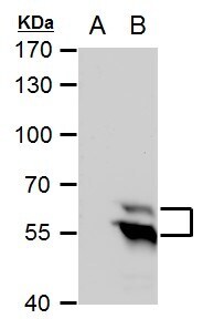
- TdT Polyclonal Antibody detects TdT protein by western blot analysis. A. 30 µg Jurkat whole cell extract. B. 30 µg Jurkat nuclear extract.7.5 % SDS-PAGE. TdT Polyclonal Antibody (Product # PA5-28880) dilution: 1:500.
- Submitted by
- Invitrogen Antibodies (provider)
- Main image
- Experimental details
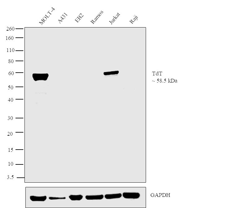
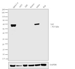
- Western blot analysis was performed on Whole cell extracts (30 µg lysate) of MOLT-4 (Lane 1), A431 (Lane 2), EB2 (Lane 3) Ramos (Lane 4), Jurkat (Lane 5) and Raji (Lane 6). The blot was probed with Anti-TdT Polyclonal Antibody (Product # PA5-28880, 1:1000 dilution) and detected by chemiluminescence using Goat anti-Rabbit IgG (H+L) Superclonal™ Secondary Antibody, HRP conjugate (Product # A27036, 0.25 µg/mL, 1:4000 dilution). A 58.5 kDa band corresponding to TdT was observed across the cell lines positive for TdT (Lanes 1 and 5), while this band was absent in cell lines which are reported negative for TdT expression (Lanes 2-4, 6).