Explore
 Validate
 Learn
Learn Western blot
 Immunohistochemistry
Antibody data
- Antibody Data
- Antigen structure
- References [0]
- Comments [0]
- Validations
- Western blot [1]
Submit
Validation data
Reference
Comment
Report error
- Product number
- PB10084 - Provider product page

- Provider
- Boster Biological Technology
- Product name
- Anti-PML Protein Antibody Picoband™
- Antibody type
- Polyclonal
- Description
- Polyclonal antibody for PML detection. Host: Rabbit.Size: 100μg/vial. Tested applications: IHC-P. Reactive species: Human. PML information: Molecular Weight: 97551 MW; Subcellular Localization: Nucleus. Nucleus, nucleoplasm. Cytoplasm. Nucleus, PML body . Nucleus, nucleolus. Endoplasmic reticulum membrane ; Peripheral membrane protein ; Cytoplasmic side . Early endosome membrane; Peripheral membrane protein; Cytoplasmic side. Isoform PML-1 can shuttle between the nucleus and cytoplasm. Isoform PML-2, isoform PML-3, isoform PML-4, isoform PML-5 and isoform PML-6 are nuclear isoforms whereas isoform PML-7 and isoform PML-14 lacking the nuclear localization signal are cytoplasmic isoforms. Detected in the nucleolus after DNA damage. Acetylation at Lys-487 is essential for its nuclear localization. Within the nucleus, most of PML is expressed in the diffuse nuclear fraction of the nucleoplasm and only a small fraction is found in the matrix-associated nuclear bodies (PML-NBs). The transfer of PML from the nucleoplasm to PML- NBs depends on its phosphorylation and sumoylation. The B1 box and the RING finger are also required for the localization in PML-NBs. Also found in specific membrane structures termed mitochondria- associated membranes (MAMs) which connect the endoplasmic reticulum (ER) and the mitochondria. Sequestered in the cytoplasm by interaction with rabies virus phosphoprotein.
- Reactivity
- Human
- Host
- Rabbit
- Vial size
- 100μg/vial
- Concentration
- Add 0.2ml of distilled water will yield a concentration of 500ug/ml.
- Storage
- At -20°C for one year. After reconstitution, at 4°C for one month. It can also be aliquoted and stored frozen at -20°C for a longer time. Avoid repeated freezing and thawing.
- Handling
- Add 0.2ml of distilled water will yield a concentration of 500ug/ml.
No comments: Submit comment
Supportive validation
- Submitted by
- Boster Biological Technology (provider)
- Main image

- Experimental details
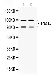
- Western blot analysis of PML using anti-PML antibody (PB10084). Electrophoresis was performed on a 5-20% SDS-PAGE gel at 70V (Stacking gel) / 90V (Resolving gel) for 2-3 hours. The sample well of each lane was loaded with 50ug of sample under reducing conditions. Lane 1: SW620 whole cell lysates, Lane 2: U20s whole cell lysates. After Electrophoresis, proteins were transferred to a Nitrocellulose membrane at 150mA for 50-90 minutes. Blocked the membrane with 5% Non-fat Milk/ TBS for 1.5 hour at RT. The membrane was incubated with rabbit anti-PML antigen affinity purified polyclonal antibody (Catalog # PB10084) at 0.5 μg/mL overnight at 4°C, then washed with TBS-0.1%Tween 3 times with 5 minutes each and probed with a goat anti-rabbit IgG-HRP secondary antibody at a dilution of 1:10000 for 1.5 hour at RT. The signal is developed using an Enhanced Chemiluminescent detection (ECL) kit (Catalog # EK1002) with Tanon 5200 system. A specific band was detected for PML at approximately 72KD, 90KD. The expected band size for PML is at 97KD.
- Additional image