Explore
 Validate
 Learn
Learn Western blot
Antibody data
- Antibody Data
- Antigen structure
- References [0]
- Comments [0]
- Validations
- Western blot [2]
Submit
Validation data
Reference
Comment
Report error
- Product number
- PA5-22754 - Provider product page

- Provider
- Invitrogen Antibodies
- Product name
- 53BP1 Polyclonal Antibody, DyLight™ 488
- Antibody type
- Polyclonal
- Antigen
- Other
- Description
- Human has been tested in WB, IHC-P and ICC/IF, mouse and goat have only been tested in ICC/IF.
- Conjugate
- Green dye
- Concentration
- 0.62 mg/mL
No comments: Submit comment
Supportive validation
- Submitted by
- Invitrogen Antibodies (provider)
- Main image

- Experimental details
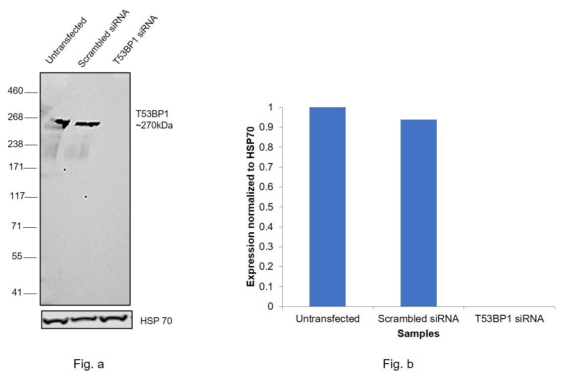
- Knockdown of TP53-binding protein 1 was achieved by transfecting HeLa with TP53-binding protein 1 specific siRNAs (Silencer® select Product # s14315, s14314). Western Blot analysis (Fig. a) was performed using Nuclear enriched extracts from the TP53-binding protein 1 knockdown cells (lane 3), non-targeting scrambled siRNA transfected cells (lane 2) and untransfected cells (lane 1). The Blot was probed with 53BP1 Polyclonal Antibody, DyLight 488 (Product # PA5-22754, 1:5000 ) and Goat anti-Rabbit IgG (H+L) Superclonal™ Recombinant Secondary Antibody, HRP (Product # A27036, 1:4000). Densitometric analysis of this western Blot is shown in histogram (Fig. b). Decrease in signal upon siRNA mediated knock down confirms that antibody is specific to TP53-binding protein 1.A streak like pattern was observed in the positive cell lines as expected of 53BP1 target.
- Conjugate
- Green dye
- Submitted by
- Invitrogen Antibodies (provider)
- Main image

- Experimental details
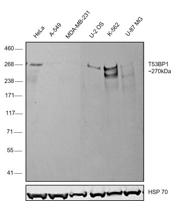
- Western Blot was performed using Anti-53BP1 Polyclonal Antibody, DyLight 488 (Product # PA5-22754) and a ~270 kDa band corresponding to TP53-binding protein 1 was observed across cell lines tested . Nuclear enriched extracts (30 µg lysate) of HeLa (Lane 1), A549 (Lane 2), MDA-MB-231 (Lane 3), U-2 OS (Lane 4), K-562 (Lane 5), U-87 MG (Lane 6) were electrophoresed using NuPAGE™ 3-8% Tris-Acetate Protein Gel (Product # EA0378BOX). Resolved proteins were then transferred onto a Nitrocellulose membrane (Product # LC2002) by iBlot® 2 Dry Blotting System (Product # IB21001). The Blot was probed with the primary antibody (1:5000) and detected by chemiluminescence with Goat anti-Rabbit IgG (H+L) Superclonal™ Recombinant Secondary Antibody, HRP (Product # A27036, 1:4000) using the iBright FL 1000 (Product # A32752). Chemiluminescent detection was performed using Novex® ECL Chemiluminescent Substrate Reagent Kit (Product # WP20005).A streak like pattern was observed in the positive cell lines as expected of 53BP1 target.
- Conjugate
- Green dye