 Explore
 Validate
 Learn
Learn Western blot
Antibody data
- Antibody Data
- Antigen structure
- References [0]
- Comments [0]
- Validations
- Western blot [4]
- Immunocytochemistry [1]
- Immunohistochemistry [1]
Submit
Validation data
Reference
Comment
Report error
- Product number
- PA5-34675 - Provider product page

- Provider
- Invitrogen Antibodies
- Product name
- 53BP1 Polyclonal Antibody
- Antibody type
- Polyclonal
- Antigen
- Recombinant protein fragment
- Description
- Recommended positive controls: 293T, HeLa, Neuro 2A, C8D30, NIH-3T3.
- Concentration
- 1 mg/mL
No comments: Submit comment
Supportive validation
- Submitted by
- Invitrogen Antibodies (provider)
- Main image
- Experimental details
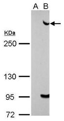
- Western blot analysis of 53BP1 using A) 30 µg 293T whole cell lysate and B) 30 whole cell lysate of human 53BP1-transfected 293T cells. Samples were loaded onto a 5% SDS-PAGE gel and probed with a 53BP1 polyclonal antibody (Product # PA5-34675) at a dilution of 1:5000.

- Submitted by
- Invitrogen Antibodies (provider)
- Main image

- Experimental details
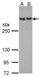
- 53BP1 Polyclonal Antibody detects TP53BP1 protein by Western blot analysis. A. 30 µg 293T whole cell lysate/extract. B. 30 µg HeLa whole cell lysate/extract.5 % SDS-PAGE.53BP1 Polyclonal Antibody (Product # PA5-34675) dilution: 1:500.

- Submitted by
- Invitrogen Antibodies (provider)
- Main image

- Experimental details
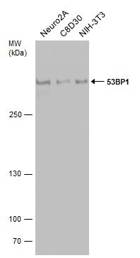
- Western Blot analysis of 53BP1 was performed by separating 30 µg of various whole cell extracts by 5% SDS-PAGE. Proteins were transferred to a membrane and probed with a 53BP1 Polyclonal Antibody (Product # PA5-34675) at a dilution of 1:500.
- Submitted by
- Invitrogen Antibodies (provider)
- Main image
- Experimental details
- 53BP1 Polyclonal Antibody detects TP53BP1 protein by Western blot analysis. A. 30 µg 293T whole cell lysate/extract. B. 30 µg HeLa whole cell lysate/extract.5 % SDS-PAGE.53BP1 Polyclonal Antibody (Product # PA5-34675) dilution: 1:500.

Supportive validation
- Submitted by
- Invitrogen Antibodies (provider)
- Main image

- Experimental details
- Immunocytochemistry-Immunofluorescence analysis of 53BP1 was performed in HeLa cells fixed in 4% paraformaldehyde at RT for 15 min. Green: 53BP1 Polyclonal Antibody (Product # PA5-34675) diluted at 1:500. Red: alpha Tubulin, a cytoskeleton marker.

Supportive validation
- Submitted by
- Invitrogen Antibodies (provider)
- Main image
- Experimental details
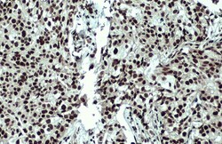
- Immunohistochemistry (Paraffin) analysis of 53BP1 was performed in paraffin-embedded human breast carcinoma tissue using 53BP1 Polyclonal Antibody (Product # PA5-34675) at a dilution of 1:200. Antigen Retrieval: Citrate buffer, pH 6.0, 15 min.
