Explore
 Validate
 Learn
Learn Western blot
Antibody data
- Antibody Data
- Antigen structure
- References [3]
- Comments [0]
- Validations
- Western blot [1]
- Immunocytochemistry [2]
- Flow cytometry [1]
Submit
Validation data
Reference
Comment
Report error
- Product number
- PAB12506 - Provider product page

- Provider
- Abnova Corporation
- Proper citation
- Abnova Corporation Cat#PAB12506, RRID:AB_10552555
- Product name
- TP53BP1 polyclonal antibody
- Antibody type
- Polyclonal
- Description
- Rabbit polyclonal antibody raised against partial recombinant TP53BP1.
- Isotype
- IgG
- Storage
- Store at -20°C.Aliquot to avoid repeated freezing and thawing.
Submitted references Cell cycle control of telomere protection and NHEJ revealed by a ts mutation in the DNA-binding domain of TRF2.
Recent expansion of the telomeric complex in rodents: Two distinct POT1 proteins protect mouse telomeres.
A pathway of double-strand break rejoining dependent upon ATM, Artemis, and proteins locating to gamma-H2AX foci.
Konishi A, de Lange T
Genes & development 2008 May 1;22(9):1221-30
Genes & development 2008 May 1;22(9):1221-30
Recent expansion of the telomeric complex in rodents: Two distinct POT1 proteins protect mouse telomeres.
Hockemeyer D, Daniels JP, Takai H, de Lange T
Cell 2006 Jul 14;126(1):63-77
Cell 2006 Jul 14;126(1):63-77
A pathway of double-strand break rejoining dependent upon ATM, Artemis, and proteins locating to gamma-H2AX foci.
Riballo E, Kühne M, Rief N, Doherty A, Smith GC, Recio MJ, Reis C, Dahm K, Fricke A, Krempler A, Parker AR, Jackson SP, Gennery A, Jeggo PA, Löbrich M
Molecular cell 2004 Dec 3;16(5):715-24
Molecular cell 2004 Dec 3;16(5):715-24
No comments: Submit comment
Supportive validation
- Submitted by
- Abnova Corporation (provider)
- Main image

- Experimental details
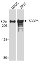
- Western blot analysis of TP53BP1 by TP53BP1 polyclonal antibody (Cat # PAB12506). Western blot Sample : Whole cell lysate (20 ug /lane) from U-2 OS or 293T cells.
Supportive validation
- Submitted by
- Abnova Corporation (provider)
- Main image
- Experimental details
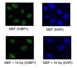
- TP53BP1 foci in proliferating MEF; both normal and exposed to 10 Gy of IR and double-color immunofluorescence stained after 2 hours. TP53BP1 stained with TP53BP1 polyclonal antibody (Cat #PAB12506).
- Validation comment
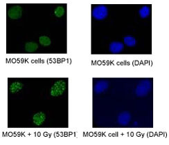
- Immunofluorescence
- Submitted by
- Abnova Corporation (provider)
- Main image
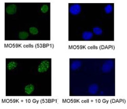
- Experimental details
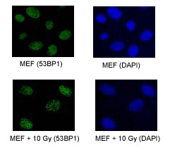
- TP53BP1 foci in proliferating M059K cells were stained with TP53BP1 polyclonal antibody (Cat # PAB12506) ; both normal and exposed to 10 Gy of IR and double-color immunofluorescence stained after 2 hours.
- Validation comment
- Immunofluorescence
Supportive validation
- Submitted by
- Abnova Corporation (provider)
- Main image
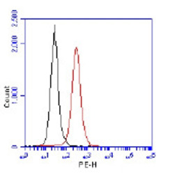
- Experimental details
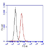
- Flow cytometric detection of TP53BP1 using TP53BP1 polyclonal antibody (Cat # PAB12506). 1 million Jurkat cells were fixed, permeabilized, and stained with TP53BP1 polyclonal antibody (Cat # PAB12506) in a 150 mcl reaction. Isotype control (Black), TP53BP1 (red).
- Validation comment
- Flow Cytometry