 Explore
 Validate
 Learn
Learn Western blot
 Immunocytochemistry
Antibody data
- Antibody Data
- Antigen structure
- References [1]
- Comments [0]
- Validations
- Western blot [1]
- Immunohistochemistry [1]
- Flow cytometry [2]
Submit
Validation data
Reference
Comment
Report error
- Product number
- NBP2-54753 - Provider product page

- Provider
- Novus Biologicals
- Product name
- Rabbit Monoclonal 53BP1 Antibody
- Antibody type
- Monoclonal
- Description
- Protein A or G purified.
- Reactivity
- Human, Mouse
- Host
- Rabbit
- Isotype
- IgG
- Vial size
- 0.1 mg
- Concentration
- 1.0 mg/ml
- Storage
- Store at 4C short term. Aliquot and store at -20C long term. Avoid freeze-thaw cycles.
Submitted references USP14 regulates DNA damage repair by targeting RNF168-dependent ubiquitination.
Sharma A, Alswillah T, Singh K, Chatterjee P, Willard B, Venere M, Summers MK, Almasan A
Autophagy 2018;14(11):1976-1990
Autophagy 2018;14(11):1976-1990
No comments: Submit comment
Supportive validation
- Submitted by
- Novus Biologicals (provider)
- Main image
- Experimental details
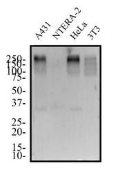
- Western Blot: 53BP1 Antibody (1285C) [NBP2-54753] - Total protein from human A431, NTERA-2, HeLa and mouse 3T3 cell lines was separated on a 12% gel by SDS-PAGE, transferred to PVDF membrane and blocked in 5% non-fat milk in TBST. The membrane was probed with 1.0 ug/ml 53BP1 Antibody in block solution and detected with an anti-rabbit HRP secondary antibody using chemiluminescence. The observed molecular weight is shown on this gel at ~250 kDa and the theoretical molecular weight of the whole endogenous protein is 214 kDa.

Supportive validation
- Submitted by
- Novus Biologicals (provider)
- Main image

- Experimental details
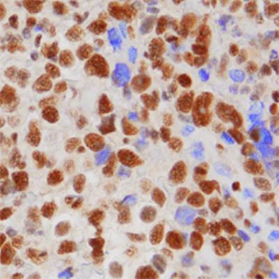
- Immunohistochemistry-Paraffin: 53BP1 Antibody (1285C) [NBP2-54753] - 53BP1 was detected in paraffin-embedded sections of human cervical cancer tissue using Rabbit Anti-Human 53BP1 Monoclonal Antibody (clone 1285C) at 1 µg/mL for 1 hour at room temperature followed by incubation with the Anti-Rabbit IgG VisUCyte™ HRP Polymer Antibody (Catalog # VC003). Tissue was stained using DAB (brown) and counterstained with hematoxylin (blue). Specific staining was localized to nuclei.
Supportive validation
- Submitted by
- Novus Biologicals (provider)
- Main image

- Experimental details
- Flow (Intracellular): 53BP1 Antibody (1285C) [NBP2-54753] - An intracellular stain was performed on HeLa Cells with 53BP1 (1285C) antibody (Catalog #NBP2-54753) (blue) and a matched isotype control MAB1050 (orange). Cells were fixed with 4% paraformaldehyde, following fixation, cells were permeabilized with 0.1% saponin. Cells were incubated in an antibody dilution of 1 ug/mL for 30 minutes at room temperature, followed by rabbit IgG APC-conjugated secondary antibody (F0111, R&D Systems).

- Submitted by
- Novus Biologicals (provider)
- Main image

- Experimental details
- Flow (Intracellular): 53BP1 Antibody (1285C) [NBP2-54753] - An intracellular stain was performed on HeLa cells with 53BP1 [1285C] Antibody (Catalog #NBP2-54753AF647) (blue) and a matched isotype control (orange). Cells were fixed with 4% PFA and then permeabilized with 0.1% saponin. Cells were incubated in an antibody dilution of 2.5 ug/mL for 30 minutes at room temperature. Both antibodies were conjugated to Alexa Fluor 647. Image using the Alexa Fluor 647 format of this antibody.
