Explore
 Validate
 Learn
LearnPA1-16565
antibody from Invitrogen Antibodies
Targeting: TP53BP1
53BP1, p202, TDRD30
 Western blot
 Immunocytochemistry Immunohistochemistry Flow cytometry
 Chromatin Immunoprecipitation Other assay
Antibody data
- Antibody Data
- Antigen structure
- References [2]
- Comments [0]
- Validations
- Immunocytochemistry [4]
- Immunohistochemistry [2]
- Flow cytometry [2]
- Other assay [1]
Submit
Validation data
Reference
Comment
Report error
- Product number
- PA1-16565 - Provider product page

- Provider
- Invitrogen Antibodies
- Product name
- 53BP1 Polyclonal Antibody
- Antibody type
- Polyclonal
- Antigen
- Synthetic peptide
- Description
- Human has been tested in WB, IHC-P and ICC/IF, mouse and goat have only been tested in ICC/IF. Suggested positive control: U205, 293T, MO59K cell lysates and MEF lysates. Predicted cross-reactivity based on sequence identity: Chimpanzee/Gorilla/Orangutan (96%), Gibbon (94%), Marmoset (92%), Feline/Porcine/Rabbit/Sheep (90%).
- Reactivity
- Human, Mouse, Rat, Bovine, Canine, Chicken/Avian, Drosophila, Goat, Porcine, Rabbit
- Host
- Rabbit
- Isotype
- IgG
- Vial size
- 100 μL
- Concentration
- 1 mg/mL
- Storage
- Store at 4°C short term. For long term storage, store at -20°C, avoiding freeze/thaw cycles.
Submitted references Effect of Ionizing Radiation from Computed Tomography on Differentiation of Human Embryonic Stem Cells into Neural Precursors.
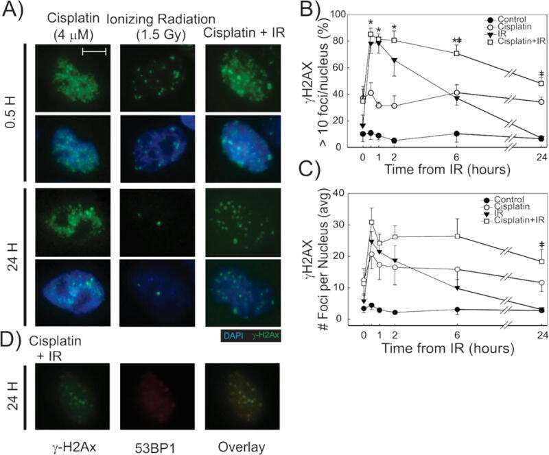
DNA damage response (DDR) pathway engagement in cisplatin radiosensitization of non-small cell lung cancer.
Hanu C, Loeliger BW, Panyutin IV, Maass-Moreno R, Wakim P, Pritchard WF, Neumann RD, Panyutin IG
International journal of molecular sciences 2019 Aug 10;20(16)
International journal of molecular sciences 2019 Aug 10;20(16)
DNA damage response (DDR) pathway engagement in cisplatin radiosensitization of non-small cell lung cancer.
Sears CR, Cooney SA, Chin-Sinex H, Mendonca MS, Turchi JJ
DNA repair 2016 Apr;40:35-46
DNA repair 2016 Apr;40:35-46
No comments: Submit comment
Supportive validation
- Submitted by
- Invitrogen Antibodies (provider)
- Main image

- Experimental details
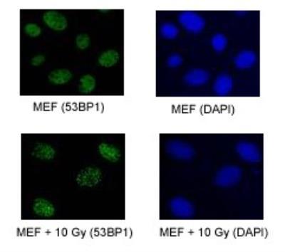
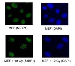
- Immunocytochemistry analysis of 53BP1 in proliferating MEFs. Samples were incubated in 53BP1 polyclonal antibody (Product # PA1-16565). Upper Panel: 53BP1 foci in proliferating MEFs. Lower Panel: 53BP1 foci in proliferating MEFs exposed to 10 Gy of IR.
- Submitted by
- Invitrogen Antibodies (provider)
- Main image
- Experimental details
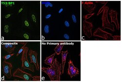
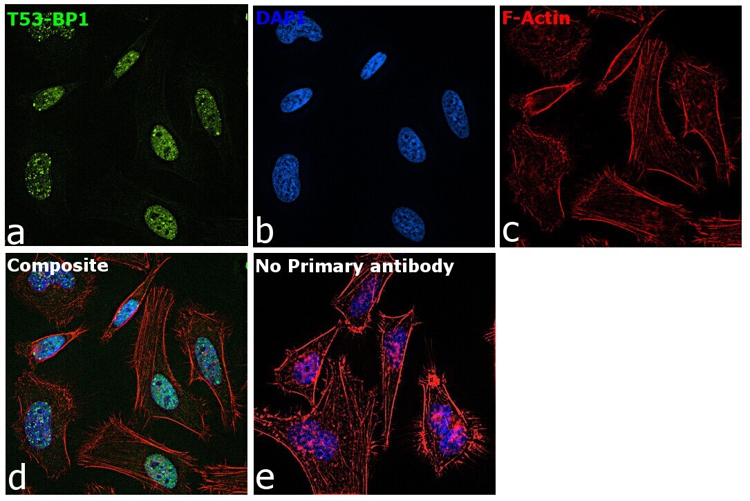
- Immunofluorescence analysis of TP53-binding protein 1 was performed using 70% confluent log phase HeLa cells. The cells were fixed with 4% paraformaldehyde for 10 minutes, permeabilized with 0.1% Triton™ X-100 for 15 minutes, and blocked with 2% BSA for 45 minutes at room temperature. The cells were labeled with 53BP1 Polyclonal Antibody (Product # PA1-16565) at 1:1000 in 0.1% BSA, incubated at 4 degree celsius overnight and then labeled with Donkey anti-Rabbit IgG (H+L) Highly Cross-Adsorbed Secondary Antibody, Alexa Fluor Plus 488 (Product # A32790), (1:2000), for 45 minutes at room temperature (Panel a: Green). Nuclei (Panel b:Blue) were stained with ProLong™ Diamond Antifade Mountant with DAPI (Product # P36962). F-actin (Panel c: Red) was stained with Rhodamine Phalloidin (Product # R415, 1:300). Panel d represents the merged image showing Nuclear localization. Panel e represents control cells with no primary antibody to assess background. The images were captured at 60X magnification.

- Submitted by
- Invitrogen Antibodies (provider)
- Main image
- Experimental details
- Immunocytochemistry analysis of 53BP1 in proliferating MEFs. Samples were incubated in 53BP1 polyclonal antibody (Product # PA1-16565). Upper Panel: 53BP1 foci in proliferating MEFs. Lower Panel: 53BP1 foci in proliferating MEFs exposed to 10 Gy of IR.

- Submitted by
- Invitrogen Antibodies (provider)
- Main image

- Experimental details
- Immunofluorescence analysis of TP53-binding protein 1 was performed using 70% confluent log phase HeLa cells. The cells were fixed with 4% paraformaldehyde for 10 minutes, permeabilized with 0.1% Triton™ X-100 for 15 minutes, and blocked with 2% BSA for 45 minutes at room temperature. The cells were labeled with 53BP1 Polyclonal Antibody (Product # PA1-16565) at 1:1000 in 0.1% BSA, incubated at 4 degree celsius overnight and then labeled with Donkey anti-Rabbit IgG (H+L) Highly Cross-Adsorbed Secondary Antibody, Alexa Fluor Plus 488 (Product # A32790), (1:2000), for 45 minutes at room temperature (Panel a: Green). Nuclei (Panel b:Blue) were stained with ProLong™ Diamond Antifade Mountant with DAPI (Product # P36962). F-actin (Panel c: Red) was stained with Rhodamine Phalloidin (Product # R415, 1:300). Panel d represents the merged image showing Nuclear localization. Panel e represents control cells with no primary antibody to assess background. The images were captured at 60X magnification.
Supportive validation
- Submitted by
- Invitrogen Antibodies (provider)
- Main image
- Experimental details
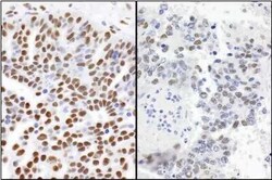
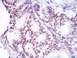
- Immunohistochemical analysis of 53BP1 in formalin-fixed paraffin-embedded sections of human ovarian carcinoma (left) and mouse teratoma (right). Samples were incubated in 53BP1 polyclonal antibody (Product # PA1-16565) using a dilution of 1:1000. Detection: DAB.
- Submitted by
- Invitrogen Antibodies (provider)
- Main image
- Experimental details
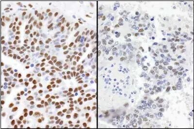
- Immunohistochemical analysis of 53BP1 in human colon cancer. Samples were incubated in 53BP1 polyclonal antibody (Product # PA1-16565) followed by DAB with hematoxylin counterstain.

Supportive validation
- Submitted by
- Invitrogen Antibodies (provider)
- Main image
- Experimental details
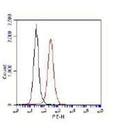
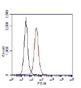
- Flow cytometric detection of 53BP1. 1 million Jurkat cells were fixed, permeabilized, and stained with 1.5 µg/mL anti-53BP1 (Product # PA1-16565) in a 150 µl reaction. Isotype control (black), anti-53BP1 (red).
- Submitted by
- Invitrogen Antibodies (provider)
- Main image
- Experimental details
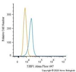
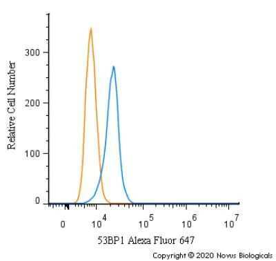
- Flow cytometry of 53BP1 in A431 cells. Samples were incubated in 53BP1 polyclonal antibody (Product # PA1-16565) using a dilution of 2.5 µg/mL for 30 minutes at room temperature. Antibody (blue) and a matched isotype control (orange). Cells were fixed with 4% PFA and then permeabilized with 0.1% saponin. Both antibodies were conjugated to Alexa Fluor 647.
Supportive validation
- Submitted by
- Invitrogen Antibodies (provider)
- Main image

- Experimental details
- NULL