 Explore
 Validate
 Learn
LearnPA1-16566
antibody from Invitrogen Antibodies
Targeting: TP53BP1
53BP1, p202, TDRD30
 Western blot
 Immunocytochemistry Immunohistochemistry Flow cytometry
 Chromatin Immunoprecipitation Other assay
Antibody data
- Antibody Data
- Antigen structure
- References [5]
- Comments [0]
- Validations
- Immunocytochemistry [2]
- Immunohistochemistry [1]
- Flow cytometry [2]
- Other assay [1]
Submit
Validation data
Reference
Comment
Report error
- Product number
- PA1-16566 - Provider product page

- Provider
- Invitrogen Antibodies
- Product name
- 53BP1 Polyclonal Antibody
- Antibody type
- Polyclonal
- Antigen
- Other
- Description
- Human samples have been tested in Western Blot and ICC/IF, and mouse samples have been tested in ICC/IF only. Suggested positive control: U205, 293T, and MO59K cell lysates and MEF lysates. Predicted cross-reactivity based on sequence identity: Chimpazee/Equine/Feline/Gibbon/Gorilla/Hamster/Marmoset/Orangutan/Panda/Porcine/Rabbit/Sheep (100%).
- Reactivity
- Human, Mouse, Rat, Bovine, Canine, Drosophila
- Host
- Rabbit
- Isotype
- IgG
- Vial size
- 100 μL
- Concentration
- 1 mg/mL
- Storage
- Store at 4°C short term. For long term storage, store at -20°C, avoiding freeze/thaw cycles.
Submitted references Loss of CENP-I Impairs Homologous Recombination and Sensitizes Cells to PARP1 Inhibition.
Multi-omics analyses of radiation survivors identify radioprotective microbes and metabolites.
Involvement of POLA2 in Double Strand Break Repair and Genotoxic Stress.
Increased DNA double-strand break was associated with downregulation of repair and upregulation of apoptotic factors in rat hippocampus after alcohol exposure.
Exposure to heavy ion radiation induces persistent oxidative stress in mouse intestine.
Dang TT, Morales JC
Cancers 2021 Jun 26;13(13)
Cancers 2021 Jun 26;13(13)
Multi-omics analyses of radiation survivors identify radioprotective microbes and metabolites.
Guo H, Chou WC, Lai Y, Liang K, Tam JW, Brickey WJ, Chen L, Montgomery ND, Li X, Bohannon LM, Sung AD, Chao NJ, Peled JU, Gomes ALC, van den Brink MRM, French MJ, Macintyre AN, Sempowski GD, Tan X, Sartor RB, Lu K, Ting JPY
Science (New York, N.Y.) 2020 Oct 30;370(6516)
Science (New York, N.Y.) 2020 Oct 30;370(6516)
Involvement of POLA2 in Double Strand Break Repair and Genotoxic Stress.
Dang TT, Morales JC
International journal of molecular sciences 2020 Jun 15;21(12)
International journal of molecular sciences 2020 Jun 15;21(12)
Increased DNA double-strand break was associated with downregulation of repair and upregulation of apoptotic factors in rat hippocampus after alcohol exposure.
Suman S, Kumar S, N'Gouemo P, Datta K
Alcohol (Fayetteville, N.Y.) 2016 Aug;54:45-50
Alcohol (Fayetteville, N.Y.) 2016 Aug;54:45-50
Exposure to heavy ion radiation induces persistent oxidative stress in mouse intestine.
Datta K, Suman S, Kallakury BV, Fornace AJ Jr
PloS one 2012;7(8):e42224
PloS one 2012;7(8):e42224
No comments: Submit comment
Supportive validation
- Submitted by
- Invitrogen Antibodies (provider)
- Main image
- Experimental details
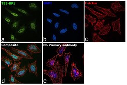
- Immunofluorescence analysis of TP53-binding protein 1 was performed using 70% confluent log phase HeLa cells. The cells were fixed with 4% paraformaldehyde for 10 minutes, permeabilized with 0.1% Triton™ X-100 for 15 minutes, and blocked with 2% BSA for 45 minutes at room temperature. The cells were labeled with 53BP1 Polyclonal Antibody (Product # PA1-16566) at 1:100 dilution in 0.1% BSA, incubated at 4 degree celsius overnight and then labeled with Donkey anti-Rabbit IgG (H+L) Highly Cross-Adsorbed Secondary Antibody, Alexa Fluor Plus 488 (Product # A32790), (1:2000), for 45 minutes at room temperature (Panel a: Green). Nuclei (Panel b:Blue) were stained with ProLong™ Diamond Antifade Mountant with DAPI (Product # P36962). F-actin (Panel c: Red) was stained with Rhodamine Phalloidin (Product # R415, 1:300). Panel d represents the merged image showing Nuclear localization. Panel e represents control cells with no primary antibody to assess background. The images were captured at 60X magnification.

- Submitted by
- Invitrogen Antibodies (provider)
- Main image

- Experimental details
- Immunofluorescence analysis of TP53-binding protein 1 was performed using 70% confluent log phase HeLa cells. The cells were fixed with 4% paraformaldehyde for 10 minutes, permeabilized with 0.1% Triton™ X-100 for 15 minutes, and blocked with 2% BSA for 45 minutes at room temperature. The cells were labeled with 53BP1 Polyclonal Antibody (Product # PA1-16566) at 1:100 dilution in 0.1% BSA, incubated at 4 degree celsius overnight and then labeled with Donkey anti-Rabbit IgG (H+L) Highly Cross-Adsorbed Secondary Antibody, Alexa Fluor Plus 488 (Product # A32790), (1:2000), for 45 minutes at room temperature (Panel a: Green). Nuclei (Panel b:Blue) were stained with ProLong™ Diamond Antifade Mountant with DAPI (Product # P36962). F-actin (Panel c: Red) was stained with Rhodamine Phalloidin (Product # R415, 1:300). Panel d represents the merged image showing Nuclear localization. Panel e represents control cells with no primary antibody to assess background. The images were captured at 60X magnification.

Supportive validation
- Submitted by
- Invitrogen Antibodies (provider)
- Main image
- Experimental details
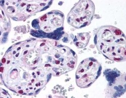
- Immunohistochemical analysis of 53BP1 in placental villi. Samples were incubated in 53BP1 polyclonal antibody (Product # PA1-16566). Antibody at 40X magnification.

Supportive validation
- Submitted by
- Invitrogen Antibodies (provider)
- Main image
- Experimental details
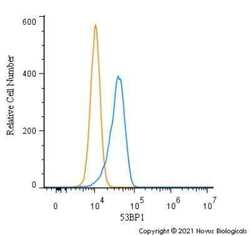
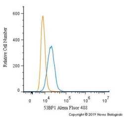
- Flow cytometry of 53BP1 in Ntera2 cells (blue) and a matched isotype control (orange). Samples were incubated in 53BP1 polyclonal antibody (Product # PA1-16566) using a dilution of 1.0 µg/mL for 30 minutes at room temperature followed by a Rabbit IgG (H+L) Cross-Adsorbed Secondary Antibody, Dylight™ 550 (Product # SA5-10033). Cells were fixed with 4% PFA and then permeabilized with 0.1% saponin.
- Submitted by
- Invitrogen Antibodies (provider)
- Main image
- Experimental details
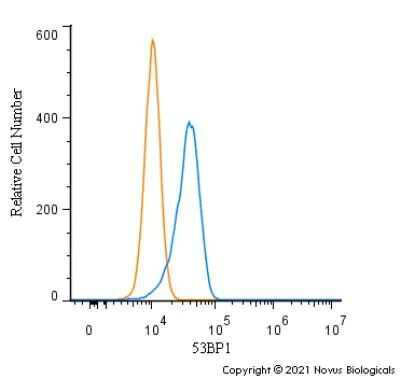
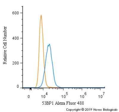
- Flow cytometry of 53BP1 in HeLa cells (blue) and a matched isotype control (orange). Samples were incubated in 53BP1 polyclonal antibody (Product # PA1-16566) using a dilution of 5 µg/mL for 30 minutes at room temperature. Cells were fixed with 4% PFA and then permeabilized with 0.1% saponin. Both antibodies were conjugated to Alexa Fluor 488.
Supportive validation
- Submitted by
- Invitrogen Antibodies (provider)
- Main image
- Experimental details
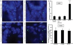
- Figure 6 Assessing 53BP1 foci and cell death in intestinal sections after gamma and 56 Fe radiation exposure. A) Intestinal sections from 6 to 8 week control, 1-year post-exposure control, 2 Gy gamma, and 1.6 Gy 56 Fe 1-year post-exposure mice were immunofluorescently stained for 53BP1. B) Quantification of 53BP1 foci in intestinal cell nuclei counterstained with DAPI. C) Quantification of TUNEL staining of intestinal sections after exposure to gamma and 56 Fe radiation. *significant compared to 6-8 wks unirradiated control; **significant compared to 1-year post-exposure unirradiated control; ***significant compared to gamma radiation.
